

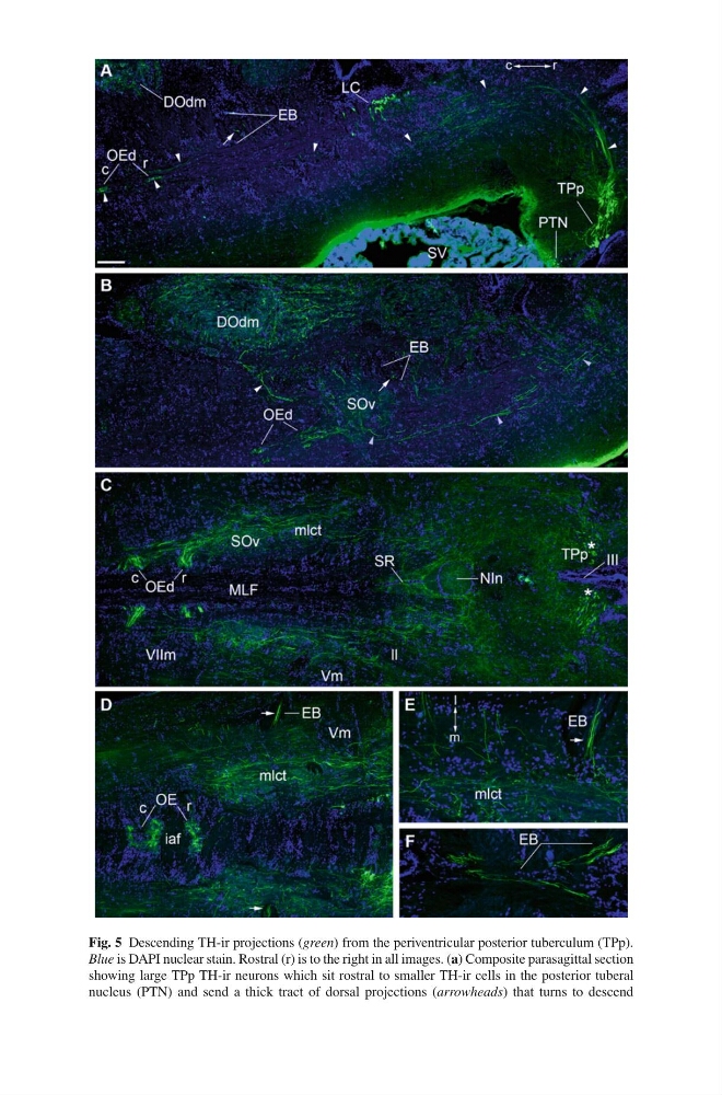
Fig. 5 Descending TH-ir projections ( green ) from the periventricular posterior tuberculum (TPp).
Blue is DAPI nuclear stain. Rostral (r) is to the right in all images. ( a ) Composite parasagittal section
showing large TPp TH-ir neurons which sit rostral to smaller TH-ir cells in the posterior tuberal
nucleus (PTN) and send a thick tract of dorsal projections ( arrowheads ) that turns to descend
Blue is DAPI nuclear stain. Rostral (r) is to the right in all images. ( a ) Composite parasagittal section
showing large TPp TH-ir neurons which sit rostral to smaller TH-ir cells in the posterior tuberal
nucleus (PTN) and send a thick tract of dorsal projections ( arrowheads ) that turns to descend