

447
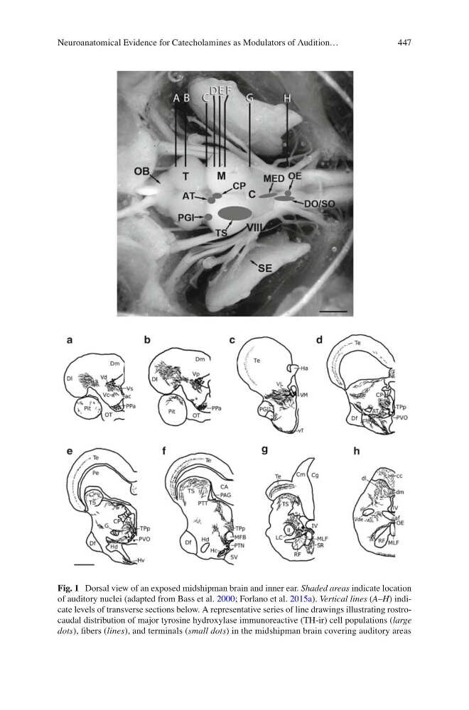
Fig. 1 Dorsal view of an exposed midshipman brain and inner ear. Shaded areas indicate location
of auditory nuclei (adapted from Bass et al. 2000 ; Forlano et al. 2015a ). Vertical lines ( A – H ) indicate levels of transverse sections below. A representative series of line drawings illustrating rostrocaudal distribution of major tyrosine hydroxylase immunoreactive (TH-ir) cell populations ( large
dots ), fi bers ( lines ), and terminals ( small dots ) in the midshipman brain covering auditory areas
Neuroanatomical Evidence for Catecholamines as Modulators of Audition…
Fig. 1 Dorsal view of an exposed midshipman brain and inner ear. Shaded areas indicate location
of auditory nuclei (adapted from Bass et al. 2000 ; Forlano et al. 2015a ). Vertical lines ( A – H ) indicate levels of transverse sections below. A representative series of line drawings illustrating rostrocaudal distribution of major tyrosine hydroxylase immunoreactive (TH-ir) cell populations ( large
dots ), fi bers ( lines ), and terminals ( small dots ) in the midshipman brain covering auditory areas
Neuroanatomical Evidence for Catecholamines as Modulators of Audition…