

429
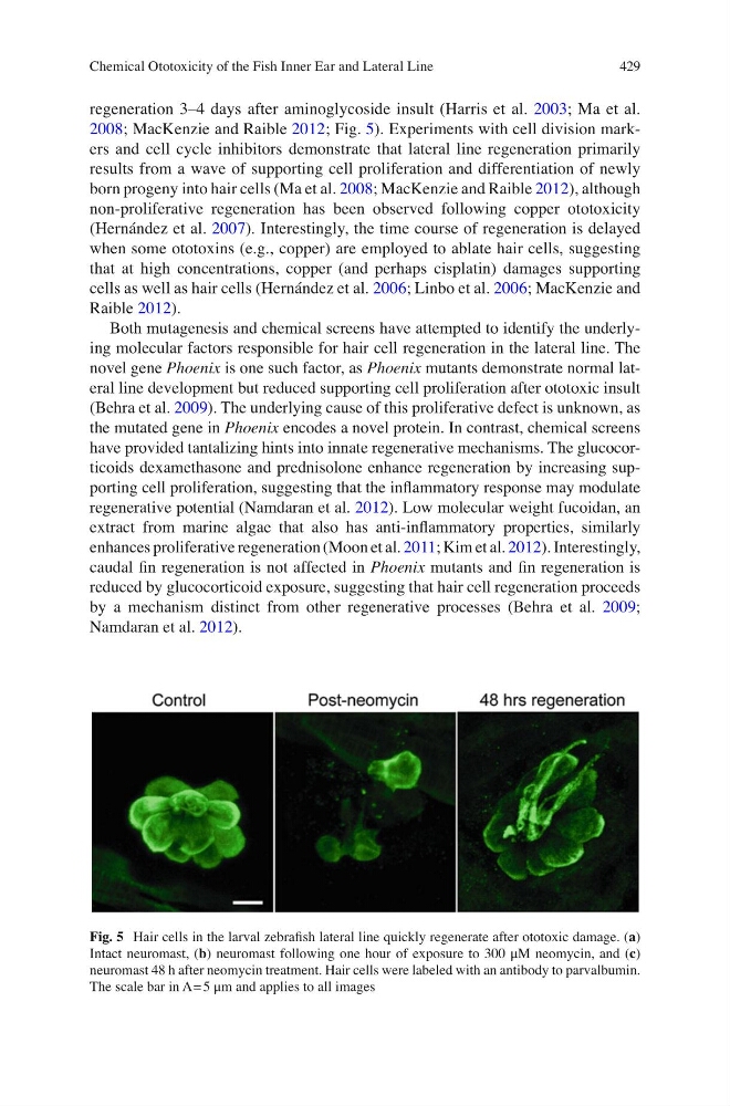
regeneration 3–4 days after aminoglycoside insult (Harris et al. 2003 ; Ma et al.
2008 ; MacKenzie and Raible 2012 ; Fig. 5 ). Experiments with cell division markers and cell cycle inhibitors demonstrate that lateral line regeneration primarily
results from a wave of supporting cell proliferation and differentiation of newly
born progeny into hair cells (Ma et al. 2008 ; MacKenzie and Raible 2012 ), although
non-proliferative regeneration has been observed following copper ototoxicity
(Hernández et al. 2007 ). Interestingly, the time course of regeneration is delayed
when some ototoxins (e.g., copper) are employed to ablate hair cells, suggesting
that at high concentrations, copper (and perhaps cisplatin) damages supporting
cells as well as hair cells (Hernández et al. 2006 ; Linbo et al. 2006 ; MacKenzie and
Raible 2012 ).
Both mutagenesis and chemical screens have attempted to identify the underlying molecular factors responsible for hair cell regeneration in the lateral line. The
novel gene Phoenix is one such factor, as Phoenix mutants demonstrate normal lateral line development but reduced supporting cell proliferation after ototoxic insult
(Behra et al. 2009 ). The underlying cause of this proliferative defect is unknown, as
the mutated gene in Phoenix encodes a novel protein. In contrast, chemical screens
have provided tantalizing hints into innate regenerative mechanisms. The glucocorticoids dexamethasone and prednisolone enhance regeneration by increasing supporting cell proliferation, suggesting that the infl ammatory response may modulate
regenerative potential (Namdaran et al. 2012 ). Low molecular weight fucoidan, an
extract from marine algae that also has anti-infl ammatory properties, similarly
enhances proliferative regeneration (Moon et al. 2011 ; Kim et al. 2012 ). Interestingly,
caudal fi n regeneration is not affected in Phoenix mutants and fi n regeneration is
reduced by glucocorticoid exposure, suggesting that hair cell regeneration proceeds
by a mechanism distinct from other regenerative processes (Behra et al. 2009 ;
Namdaran et al. 2012 ).
Fig. 5 Hair cells in the larval zebrafi sh lateral line quickly regenerate after ototoxic damage. ( a )
Intact neuromast, ( b ) neuromast following one hour of exposure to 300 μM neomycin, and ( c )
neuromast 48 h after neomycin treatment. Hair cells were labeled with an antibody to parvalbumin.
The scale bar in A = 5 μm and applies to all images
Chemical Ototoxicity of the Fish Inner Ear and Lateral Line
regeneration 3–4 days after aminoglycoside insult (Harris et al. 2003 ; Ma et al.
2008 ; MacKenzie and Raible 2012 ; Fig. 5 ). Experiments with cell division markers and cell cycle inhibitors demonstrate that lateral line regeneration primarily
results from a wave of supporting cell proliferation and differentiation of newly
born progeny into hair cells (Ma et al. 2008 ; MacKenzie and Raible 2012 ), although
non-proliferative regeneration has been observed following copper ototoxicity
(Hernández et al. 2007 ). Interestingly, the time course of regeneration is delayed
when some ototoxins (e.g., copper) are employed to ablate hair cells, suggesting
that at high concentrations, copper (and perhaps cisplatin) damages supporting
cells as well as hair cells (Hernández et al. 2006 ; Linbo et al. 2006 ; MacKenzie and
Raible 2012 ).
Both mutagenesis and chemical screens have attempted to identify the underlying molecular factors responsible for hair cell regeneration in the lateral line. The
novel gene Phoenix is one such factor, as Phoenix mutants demonstrate normal lateral line development but reduced supporting cell proliferation after ototoxic insult
(Behra et al. 2009 ). The underlying cause of this proliferative defect is unknown, as
the mutated gene in Phoenix encodes a novel protein. In contrast, chemical screens
have provided tantalizing hints into innate regenerative mechanisms. The glucocorticoids dexamethasone and prednisolone enhance regeneration by increasing supporting cell proliferation, suggesting that the infl ammatory response may modulate
regenerative potential (Namdaran et al. 2012 ). Low molecular weight fucoidan, an
extract from marine algae that also has anti-infl ammatory properties, similarly
enhances proliferative regeneration (Moon et al. 2011 ; Kim et al. 2012 ). Interestingly,
caudal fi n regeneration is not affected in Phoenix mutants and fi n regeneration is
reduced by glucocorticoid exposure, suggesting that hair cell regeneration proceeds
by a mechanism distinct from other regenerative processes (Behra et al. 2009 ;
Namdaran et al. 2012 ).
Fig. 5 Hair cells in the larval zebrafi sh lateral line quickly regenerate after ototoxic damage. ( a )
Intact neuromast, ( b ) neuromast following one hour of exposure to 300 μM neomycin, and ( c )
neuromast 48 h after neomycin treatment. Hair cells were labeled with an antibody to parvalbumin.
The scale bar in A = 5 μm and applies to all images
Chemical Ototoxicity of the Fish Inner Ear and Lateral Line