

375
bandwidth and higher auditory sensitivities was shown for the sciaenid B. chrysoura
and the cichlids E. maculatus and Paratilapia polleni (Ramcharitar et al. 2004 ;
Schulz-Mirbach et al. 2012 ).
3 Evolution of Ciliary Bundle Orientation Patterns
What was the ancestral state in the teleostean macula sacculi: the vertical pattern
or a pattern with vertical and horizontal groups? Tetrapods have only two “vertical” groups on the macula sacculi and this may also hold true for non-teleost actinopterygians as well as lungfi shes: the horizontal groups in these fi shes are
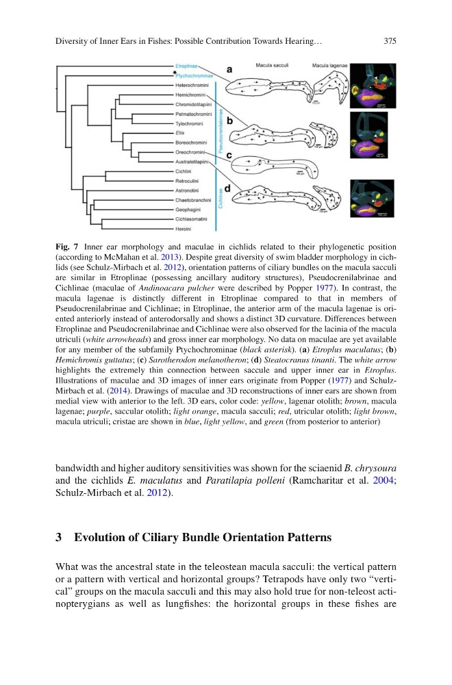
Fig. 7 Inner ear morphology and maculae in cichlids related to their phylogenetic position
(according to McMahan et al. 2013 ). Despite great diversity of swim bladder morphology in cichlids (see Schulz-Mirbach et al. 2012 ), orientation patterns of ciliary bundles on the macula sacculi
are similar in Etroplinae (possessing ancillary auditory structures), Pseudocrenilabrinae and
Cichlinae (maculae of Andinoacara pulcher were described by Popper 1977 ). In contrast, the
macula lagenae is distinctly different in Etroplinae compared to that in members of
Pseudocrenilabrinae and Cichlinae; in Etroplinae, the anterior arm of the macula lagenae is oriented anteriorly instead of anterodorsally and shows a distinct 3D curvature. Differences between
Etroplinae and Pseudocrenilabrinae and Cichlinae were also observed for the lacinia of the macula
utriculi ( white arrowheads ) and gross inner ear morphology. No data on maculae are yet available
for any member of the subfamily Ptychochrominae ( black asterisk ). ( a ) Etroplus maculatus ; ( b )
Hemichromis guttatus ; ( c ) Sarotherodon melanotheron ; ( d ) Steatocranus tinanti . The white arrow
highlights the extremely thin connection between saccule and upper inner ear in Etroplus .
Illustrations of maculae and 3D images of inner ears originate from Popper ( 1977 ) and SchulzMirbach et al. ( 2014 ). Drawings of maculae and 3D reconstructions of inner ears are shown from
medial view with anterior to the left. 3D ears, color code: yellow , lagenar otolith; brown , macula
lagenae; purple , saccular otolith; light orange , macula sacculi; red , utricular otolith; light brown ,
macula utriculi; cristae are shown in blue , light yellow , and green (from posterior to anterior)
Diversity of Inner Ears in Fishes: Possible Contribution Towards Hearing…
bandwidth and higher auditory sensitivities was shown for the sciaenid B. chrysoura
and the cichlids E. maculatus and Paratilapia polleni (Ramcharitar et al. 2004 ;
Schulz-Mirbach et al. 2012 ).
3 Evolution of Ciliary Bundle Orientation Patterns
What was the ancestral state in the teleostean macula sacculi: the vertical pattern
or a pattern with vertical and horizontal groups? Tetrapods have only two “vertical” groups on the macula sacculi and this may also hold true for non-teleost actinopterygians as well as lungfi shes: the horizontal groups in these fi shes are
Fig. 7 Inner ear morphology and maculae in cichlids related to their phylogenetic position
(according to McMahan et al. 2013 ). Despite great diversity of swim bladder morphology in cichlids (see Schulz-Mirbach et al. 2012 ), orientation patterns of ciliary bundles on the macula sacculi
are similar in Etroplinae (possessing ancillary auditory structures), Pseudocrenilabrinae and
Cichlinae (maculae of Andinoacara pulcher were described by Popper 1977 ). In contrast, the
macula lagenae is distinctly different in Etroplinae compared to that in members of
Pseudocrenilabrinae and Cichlinae; in Etroplinae, the anterior arm of the macula lagenae is oriented anteriorly instead of anterodorsally and shows a distinct 3D curvature. Differences between
Etroplinae and Pseudocrenilabrinae and Cichlinae were also observed for the lacinia of the macula
utriculi ( white arrowheads ) and gross inner ear morphology. No data on maculae are yet available
for any member of the subfamily Ptychochrominae ( black asterisk ). ( a ) Etroplus maculatus ; ( b )
Hemichromis guttatus ; ( c ) Sarotherodon melanotheron ; ( d ) Steatocranus tinanti . The white arrow
highlights the extremely thin connection between saccule and upper inner ear in Etroplus .
Illustrations of maculae and 3D images of inner ears originate from Popper ( 1977 ) and SchulzMirbach et al. ( 2014 ). Drawings of maculae and 3D reconstructions of inner ears are shown from
medial view with anterior to the left. 3D ears, color code: yellow , lagenar otolith; brown , macula
lagenae; purple , saccular otolith; light orange , macula sacculi; red , utricular otolith; light brown ,
macula utriculi; cristae are shown in blue , light yellow , and green (from posterior to anterior)
Diversity of Inner Ears in Fishes: Possible Contribution Towards Hearing…