

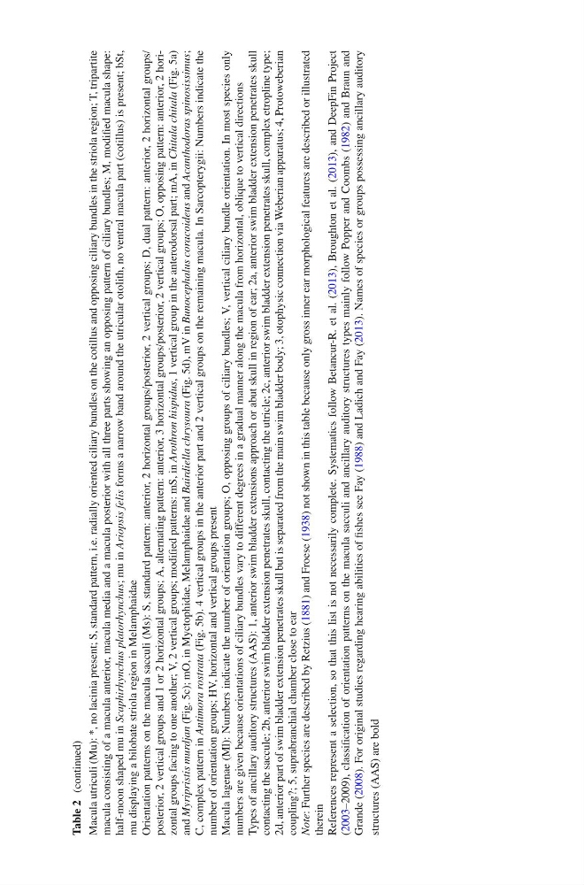
Macula utriculi (Mu): *, no lacinia present; S, standard pattern, i.e. radially oriented ciliary bundles on the cotillus and opposing ciliary bundles in the striola region; T, tripartite
macula consisting of a macula anterior, macula media and a macula posterior with all three parts showing an opposing pattern of ciliary bundles; M, modifi ed macula shape:
half-moon shaped mu in Scaphirhynchus platorhynchus
; mu in Ariopsis felis
forms a narrow band around the utricular otolith, no ventral macula part (cotillus) is present; bSt,
mu displaying a bilobate striola region in Melamphaidae
Orientation patterns on the macula sacculi (Ms): S, standard pattern: anterior, 2 horizontal groups/posterior, 2 vertical groups; D, dual pattern: anterior, 2 horizontal groups/
posterior, 2 vertical groups and 1 or 2 horizontal groups; A, alternating pattern: anterior, 3 horizontal groups/posterior, 2 vertical groups; O, opposing pattern: anterior, 2 horizontal groups facing to one another; V, 2 vertical groups; modifi ed patterns: mS, in Arothron hispidus
, 1 vertical group in the anterodorsal part; mA, in Chitala chitala
(Fig. 5a )
and Myripristis murdjan
(Fig. 5c ); mO, in Myctophidae, Melamphaidae and Bairdiella chrysoura
(Fig. 5d ), mV in Bunocephalus coracoideus
and Acanthodoras spinosissimus
;
C, complex pattern in Antimora rostrata
(Fig. 5b ). 4 vertical groups in the anterior part and 2 vertical groups on the remaining macula. In Sarcopterygii: Numbers indicate the
number of orientation groups; HV, horizontal and vertical groups present
Macula lagenae (Ml): Numbers indicate the number of orientation groups; O, opposing groups of ciliary bundles; V, vertical ciliary bundle orientation. In most species only
numbers are given because orientations of ciliary bundles vary to different degrees in a gradual manner along the macula from horizontal, oblique to vertical directions
Types of ancillary auditory structures (AAS): 1, anterior swim bladder extensions approach or abut skull in region of ear; 2a, anterior swim bladder extension penetrates skull
contacting the saccule; 2b, anterior swim bladder extension penetrates skull, contacting the utricle; 2c, anterior swim bladder extension penetrates skull, complex etropline type;
2d, anterior part of swim bladder extension penetrates skull but is separated from the main swim bladder body; 3, otophysic connection via Weberian apparatus; 4, Protoweberian
coupling?; 5, suprabranchial chamber close to ear
Note
: Further species are described by Retzius ( 1881 ) and Froese ( 1938 ) not shown in this table because only gross inner ear morphological features are described or illustrated
therein
References represent a selection, so that this list is not necessarily complete. Systematics follow Betancur-R. et al. ( 2013 ), Broughton et al. ( 2013 ), and DeepFin Project
( 2003 –2009), classifi cation of orientation patterns on the macula sacculi and ancillary auditory structures types mainly follow Popper and Coombs ( 1982 ) and Braun and
Grande ( 2008 ). For original studies regarding hearing abilities of fi shes see Fay ( 1988 ) and Ladich and Fay ( 2013 ). Names of species or groups possessing ancillary auditory
structures (AAS) are bold
Table 2
(continued)
macula consisting of a macula anterior, macula media and a macula posterior with all three parts showing an opposing pattern of ciliary bundles; M, modifi ed macula shape:
half-moon shaped mu in Scaphirhynchus platorhynchus
; mu in Ariopsis felis
forms a narrow band around the utricular otolith, no ventral macula part (cotillus) is present; bSt,
mu displaying a bilobate striola region in Melamphaidae
Orientation patterns on the macula sacculi (Ms): S, standard pattern: anterior, 2 horizontal groups/posterior, 2 vertical groups; D, dual pattern: anterior, 2 horizontal groups/
posterior, 2 vertical groups and 1 or 2 horizontal groups; A, alternating pattern: anterior, 3 horizontal groups/posterior, 2 vertical groups; O, opposing pattern: anterior, 2 horizontal groups facing to one another; V, 2 vertical groups; modifi ed patterns: mS, in Arothron hispidus
, 1 vertical group in the anterodorsal part; mA, in Chitala chitala
(Fig. 5a )
and Myripristis murdjan
(Fig. 5c ); mO, in Myctophidae, Melamphaidae and Bairdiella chrysoura
(Fig. 5d ), mV in Bunocephalus coracoideus
and Acanthodoras spinosissimus
;
C, complex pattern in Antimora rostrata
(Fig. 5b ). 4 vertical groups in the anterior part and 2 vertical groups on the remaining macula. In Sarcopterygii: Numbers indicate the
number of orientation groups; HV, horizontal and vertical groups present
Macula lagenae (Ml): Numbers indicate the number of orientation groups; O, opposing groups of ciliary bundles; V, vertical ciliary bundle orientation. In most species only
numbers are given because orientations of ciliary bundles vary to different degrees in a gradual manner along the macula from horizontal, oblique to vertical directions
Types of ancillary auditory structures (AAS): 1, anterior swim bladder extensions approach or abut skull in region of ear; 2a, anterior swim bladder extension penetrates skull
contacting the saccule; 2b, anterior swim bladder extension penetrates skull, contacting the utricle; 2c, anterior swim bladder extension penetrates skull, complex etropline type;
2d, anterior part of swim bladder extension penetrates skull but is separated from the main swim bladder body; 3, otophysic connection via Weberian apparatus; 4, Protoweberian
coupling?; 5, suprabranchial chamber close to ear
Note
: Further species are described by Retzius ( 1881 ) and Froese ( 1938 ) not shown in this table because only gross inner ear morphological features are described or illustrated
therein
References represent a selection, so that this list is not necessarily complete. Systematics follow Betancur-R. et al. ( 2013 ), Broughton et al. ( 2013 ), and DeepFin Project
( 2003 –2009), classifi cation of orientation patterns on the macula sacculi and ancillary auditory structures types mainly follow Popper and Coombs ( 1982 ) and Braun and
Grande ( 2008 ). For original studies regarding hearing abilities of fi shes see Fay ( 1988 ) and Ladich and Fay ( 2013 ). Names of species or groups possessing ancillary auditory
structures (AAS) are bold
Table 2
(continued)