

208
directional computations along with the saccule. McCormick and Wallace (2012)
conducted an elegant study in goldfish in which they were able to show that all three
otolithic endorgans send projections to identified auditory projection cells in
DON. Although the saccule contributed the most input to the auditory cells in the
goldfish DON, most often with bouton-like endings on the somata, McCormick and
Wallace found evidence that the utricle or lagena also provided input to some of
those auditory projection cells (see Table 2, McCormick and Wallace 2012). The
anatomy indicates convergence of the otolithic endorgan inputs in the dorsal regions
of the DON, but the physiological response properties of the DON projection cells
receiving those inputs are unknown. Given that the goldfish has an otophysic connection that mechanically links movement of the gas bladder to the fluids in the ear,
resulting in enhanced reception of the pressure component of sound, input from the
endorgan encoding the indirect particle motion from the gas bladder (presumed to
be the saccule in goldfish) may be parceled in some way for phase comparisons with
input from an endorgan receiving only direct particle motion.
If the organization of dDON in goldfish includes a division for phase comparisons, projections from otolithic afferent input to the dDON in a fish lacking an
otophysic connection may reveal a different organization of inputs. Tomchik and Lu
(2005) examined the central projections of afferents from all three otolithic endorgans in the “non-otophysic” sleeper goby, in which, like the goldfish, the saccule,
utricle, and lagena have overlapping frequency responses and distinct directionality.
They found inter-digitating projection sites for the three otolithic endorgans in the
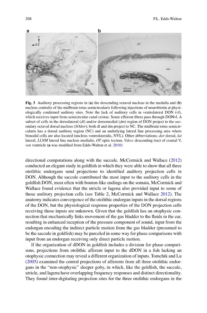
Fig. 3 Auditory processing regions in (a) the descending octaval nucleus in the medulla and (b)
nucleus centralis of the midbrain torus semicircularis following injections of neurobiotin at physiologically confirmed auditory sites. Note the lack of auditory cells in ventrolateral DON (vl),
which receives input from semicircular canal cristae. Some efferent fibers pass through DONvl. A
subset of cells in the dorsolateral (dl) and/or dorsomedial (dm) region of DON project to the secondary octaval dorsal nucleus (SOdor); both dl and dm project to NC. The midbrain torus semicircularis has a dorsal auditory region (NC) and an underlying lateral line processing area where
bimodal cells are also located (nucleus ventrolateralis, NVL). Other abbreviations: dor dorsal, lat
lateral, LLNM lateral line nucleus medialis, OT optic tectum, Vdesc descending tract of cranial V,
ven ventricle (a was modified from Edds-Walton et al. 2010)
P.L. Edds-Walton
directional computations along with the saccule. McCormick and Wallace (2012)
conducted an elegant study in goldfish in which they were able to show that all three
otolithic endorgans send projections to identified auditory projection cells in
DON. Although the saccule contributed the most input to the auditory cells in the
goldfish DON, most often with bouton-like endings on the somata, McCormick and
Wallace found evidence that the utricle or lagena also provided input to some of
those auditory projection cells (see Table 2, McCormick and Wallace 2012). The
anatomy indicates convergence of the otolithic endorgan inputs in the dorsal regions
of the DON, but the physiological response properties of the DON projection cells
receiving those inputs are unknown. Given that the goldfish has an otophysic connection that mechanically links movement of the gas bladder to the fluids in the ear,
resulting in enhanced reception of the pressure component of sound, input from the
endorgan encoding the indirect particle motion from the gas bladder (presumed to
be the saccule in goldfish) may be parceled in some way for phase comparisons with
input from an endorgan receiving only direct particle motion.
If the organization of dDON in goldfish includes a division for phase comparisons, projections from otolithic afferent input to the dDON in a fish lacking an
otophysic connection may reveal a different organization of inputs. Tomchik and Lu
(2005) examined the central projections of afferents from all three otolithic endorgans in the “non-otophysic” sleeper goby, in which, like the goldfish, the saccule,
utricle, and lagena have overlapping frequency responses and distinct directionality.
They found inter-digitating projection sites for the three otolithic endorgans in the
Fig. 3 Auditory processing regions in (a) the descending octaval nucleus in the medulla and (b)
nucleus centralis of the midbrain torus semicircularis following injections of neurobiotin at physiologically confirmed auditory sites. Note the lack of auditory cells in ventrolateral DON (vl),
which receives input from semicircular canal cristae. Some efferent fibers pass through DONvl. A
subset of cells in the dorsolateral (dl) and/or dorsomedial (dm) region of DON project to the secondary octaval dorsal nucleus (SOdor); both dl and dm project to NC. The midbrain torus semicircularis has a dorsal auditory region (NC) and an underlying lateral line processing area where
bimodal cells are also located (nucleus ventrolateralis, NVL). Other abbreviations: dor dorsal, lat
lateral, LLNM lateral line nucleus medialis, OT optic tectum, Vdesc descending tract of cranial V,
ven ventricle (a was modified from Edds-Walton et al. 2010)
P.L. Edds-Walton