

198
1 Introduction
When considering the vast work on fishes conducted by Arthur Popper and Richard
Fay, the obvious theme is a better understanding of how (and what) fish hear.
However, each took a different approach. Much of Popper’s fish research was driven
by an interest in the organization and functions of the sensory hair cells, what one
might call a “bottom-up approach.” Much of Fay’s research used psychophysical
methods to investigate and define the limits of the sense of hearing in fishes, using
a comparative approach and methods previously used for other vertebrates, particularly mammals, which one might call a “top-down approach.”
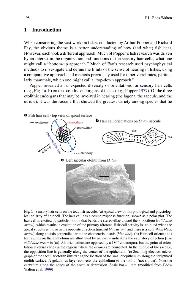
Popper revealed an unexpected diversity of orientations for sensory hair cells
(e.g., Fig. 1a, b) on the otolithic endorgans of fishes (e.g., Popper 1977). Of the three
otolithic endorgans that may be involved in hearing (the lagena, the saccule, and the
utricle), it was the saccule that showed the greatest variety among species that he
Fig. 1 Sensory hair cells on the toadfish saccule. (a) Apical view of morphological and physiological polarity of hair cell. The hair cell has a cosine response function, shown as a polar plot. The
hair cell is excited by particle motion that bends the stereovillae toward the kinocilium (solid blue
arrow), which results in excitation of the primary afferent. Hair cell activity is inhibited when the
apical structures move in the opposite direction (dashed blue arrow) and there is a null (thick black
arrow) along an axis perpendicular to the characteristic axis (blue line). (b) Hair cell orientations
for regions on the epithelium are illustrated by an arrow indicating the excitatory direction [like
solid blue arrow in (a)]. All orientations are opposed by a 180° counterpart, but the point of orientation reversal varies in the regions where the arrows are connected. In the middle of the saccule,
the opposition line is generally along the center of the epithelium. (c) Scanning electron micrograph of the saccular otolith illustrating the location of the smaller epithelium along the sculptured
otolith surface. A gelatinous layer connects the epithelium to the otolith (not shown). Note the
curvature along the edges of the saccular depression. Scale bar = 1 mm (modified from EddsWalton et al. 1999)
P.L. Edds-Walton
1 Introduction
When considering the vast work on fishes conducted by Arthur Popper and Richard
Fay, the obvious theme is a better understanding of how (and what) fish hear.
However, each took a different approach. Much of Popper’s fish research was driven
by an interest in the organization and functions of the sensory hair cells, what one
might call a “bottom-up approach.” Much of Fay’s research used psychophysical
methods to investigate and define the limits of the sense of hearing in fishes, using
a comparative approach and methods previously used for other vertebrates, particularly mammals, which one might call a “top-down approach.”
Popper revealed an unexpected diversity of orientations for sensory hair cells
(e.g., Fig. 1a, b) on the otolithic endorgans of fishes (e.g., Popper 1977). Of the three
otolithic endorgans that may be involved in hearing (the lagena, the saccule, and the
utricle), it was the saccule that showed the greatest variety among species that he
Fig. 1 Sensory hair cells on the toadfish saccule. (a) Apical view of morphological and physiological polarity of hair cell. The hair cell has a cosine response function, shown as a polar plot. The
hair cell is excited by particle motion that bends the stereovillae toward the kinocilium (solid blue
arrow), which results in excitation of the primary afferent. Hair cell activity is inhibited when the
apical structures move in the opposite direction (dashed blue arrow) and there is a null (thick black
arrow) along an axis perpendicular to the characteristic axis (blue line). (b) Hair cell orientations
for regions on the epithelium are illustrated by an arrow indicating the excitatory direction [like
solid blue arrow in (a)]. All orientations are opposed by a 180° counterpart, but the point of orientation reversal varies in the regions where the arrows are connected. In the middle of the saccule,
the opposition line is generally along the center of the epithelium. (c) Scanning electron micrograph of the saccular otolith illustrating the location of the smaller epithelium along the sculptured
otolith surface. A gelatinous layer connects the epithelium to the otolith (not shown). Note the
curvature along the edges of the saccular depression. Scale bar = 1 mm (modified from EddsWalton et al. 1999)
P.L. Edds-Walton