

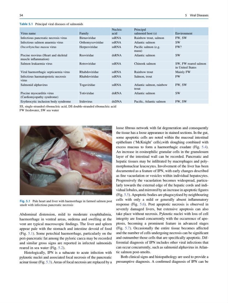
Abdominal distension, mild to moderate exophthalmia,
haemorrhage in ventral areas, oedema and swelling at the
vent are typical macroscopic findings. The liver and spleen
appear pale with the stomach and intestine devoid of food
(Fig. 5.1). Some petechial haemorrhage, particularly on the
peri-pancreatic fat among the pyloric caeca may be recorded
and similar gross signs are reported in infected salmonids
reared in sea water (Fig. 5.2).
Histologically, IPN is a subacute to acute infection with
pyknotic nuclei and associated focal necrosis of the pancreatic
acinar tissue (Fig. 5.3). Areas of focal necrosis are replaced by a
loose fibrous network with fat degeneration and consequently
the tissue has a loose appearance in stained sections. In the gut,
some apoptotic cells are noted within the mucosal intestinal
epithelium (‘McKnight’ cells),with sloughing combined with
excess mucous to form a haemorrhagic exudate (Fig. 5.4).
An increase in eosinophilic granular cells in the granulosum
layer of the intestinal wall can be recorded. Pancreatic and
hepatic tissues may be infiltrated by macrophages and polymorphonuclear leucocytes. Involvement of the liver has been
documented as a feature of IPN, with early changes described
as fine vacuolation or vesicles within individual hepatocytes.
Progressively the vacuolation becomes widespread, particularly towards the external edge of the hepatic cords and individual lobules, and mirrored by an increase in apoptotic figures
(Fig. 5.5). Apoptotic bodies are phagocytised by neighbouring
cells with only a mild or generally absent inflammatory
response (Fig. 5.6). Post apoptotic necrosis is observed in
severely damaged livers, but extensive apoptosis can also
take place without necrosis. Pyknotic nuclei with loss of cell
integrity are found concurrently with the occurrence of apoptosis, becoming a prominent feature in advanced stages
(Fig. 5.7). Occasionally the entire tissue becomes affected
and the number of cells undergoing necrosis can be significant
and outnumber those cells that are specifically apoptotic. Differential diagnosis of IPN includes other viral infections that
can occur concurrently, such as salmonid alphavirus in Atlantic salmon post-smolts.
Both clinical signs and histopathology are used to provide a
presumptive diagnosis. A confirmed diagnosis of IPN can be
Table 5.1 Principal viral diseases of salmonids
Virus name
Family
Nucleic
acid
Principal
salmonid host (s)
Environment
Infectious pancreatic necrosis virus
Birnaviridae
ssRNA
Rainbow trout, salmon
FW, SW
Infectious salmon anaemia virus
Orthomyxoviridae
ssRNA
Atlantic salmon
SW
Oncorhynchus masou virus
Herpesviridae
ssRNA
Pacific salmon (e.g.
masu)
FW?
Piscine reovirus (Heart and skeletal
muscle inflammation)
Reoviridae
dsRNA
Atlantic salmon
SW
Salmon leukaemia virus
Retroviridae
ssRNA
Chinook salmon
SW, FW reared salmon
in United States
Viral haemorrhagic septicaemia virus
Rhabdoviridae
ssRNA
Rainbow trout
Mainly FW
Infectious haematopoietic necrosis
virus
Rhabdoviridae
ssRNA
Salmon, trout
FW
Salmonid alphavirus
Togaviridae
ssRNA
Atlantic salmon, rainbow
trout
FW, SW
Piscine myocarditis virus
(Cardiomyopathy syndrome)
Totiviridae
dsRNA
Atlantic salmon
SW
Erythrocytic inclusion body syndrome
Iridovirus
dsDNA
Pacific, Atlantic salmon
FW, SW
SS, single-stranded ribonucleic acid, DS double-stranded ribonucleic acid
FW freshwater, SW sea water
Fig. 5.1 Pale heart and liver with haemorrhage in farmed salmon post
smolt with infectious pancreatic necrosis
54
5 Viral Diseases
haemorrhage in ventral areas, oedema and swelling at the
vent are typical macroscopic findings. The liver and spleen
appear pale with the stomach and intestine devoid of food
(Fig. 5.1). Some petechial haemorrhage, particularly on the
peri-pancreatic fat among the pyloric caeca may be recorded
and similar gross signs are reported in infected salmonids
reared in sea water (Fig. 5.2).
Histologically, IPN is a subacute to acute infection with
pyknotic nuclei and associated focal necrosis of the pancreatic
acinar tissue (Fig. 5.3). Areas of focal necrosis are replaced by a
loose fibrous network with fat degeneration and consequently
the tissue has a loose appearance in stained sections. In the gut,
some apoptotic cells are noted within the mucosal intestinal
epithelium (‘McKnight’ cells),with sloughing combined with
excess mucous to form a haemorrhagic exudate (Fig. 5.4).
An increase in eosinophilic granular cells in the granulosum
layer of the intestinal wall can be recorded. Pancreatic and
hepatic tissues may be infiltrated by macrophages and polymorphonuclear leucocytes. Involvement of the liver has been
documented as a feature of IPN, with early changes described
as fine vacuolation or vesicles within individual hepatocytes.
Progressively the vacuolation becomes widespread, particularly towards the external edge of the hepatic cords and individual lobules, and mirrored by an increase in apoptotic figures
(Fig. 5.5). Apoptotic bodies are phagocytised by neighbouring
cells with only a mild or generally absent inflammatory
response (Fig. 5.6). Post apoptotic necrosis is observed in
severely damaged livers, but extensive apoptosis can also
take place without necrosis. Pyknotic nuclei with loss of cell
integrity are found concurrently with the occurrence of apoptosis, becoming a prominent feature in advanced stages
(Fig. 5.7). Occasionally the entire tissue becomes affected
and the number of cells undergoing necrosis can be significant
and outnumber those cells that are specifically apoptotic. Differential diagnosis of IPN includes other viral infections that
can occur concurrently, such as salmonid alphavirus in Atlantic salmon post-smolts.
Both clinical signs and histopathology are used to provide a
presumptive diagnosis. A confirmed diagnosis of IPN can be
Table 5.1 Principal viral diseases of salmonids
Virus name
Family
Nucleic
acid
Principal
salmonid host (s)
Environment
Infectious pancreatic necrosis virus
Birnaviridae
ssRNA
Rainbow trout, salmon
FW, SW
Infectious salmon anaemia virus
Orthomyxoviridae
ssRNA
Atlantic salmon
SW
Oncorhynchus masou virus
Herpesviridae
ssRNA
Pacific salmon (e.g.
masu)
FW?
Piscine reovirus (Heart and skeletal
muscle inflammation)
Reoviridae
dsRNA
Atlantic salmon
SW
Salmon leukaemia virus
Retroviridae
ssRNA
Chinook salmon
SW, FW reared salmon
in United States
Viral haemorrhagic septicaemia virus
Rhabdoviridae
ssRNA
Rainbow trout
Mainly FW
Infectious haematopoietic necrosis
virus
Rhabdoviridae
ssRNA
Salmon, trout
FW
Salmonid alphavirus
Togaviridae
ssRNA
Atlantic salmon, rainbow
trout
FW, SW
Piscine myocarditis virus
(Cardiomyopathy syndrome)
Totiviridae
dsRNA
Atlantic salmon
SW
Erythrocytic inclusion body syndrome
Iridovirus
dsDNA
Pacific, Atlantic salmon
FW, SW
SS, single-stranded ribonucleic acid, DS double-stranded ribonucleic acid
FW freshwater, SW sea water
Fig. 5.1 Pale heart and liver with haemorrhage in farmed salmon post
smolt with infectious pancreatic necrosis
54
5 Viral Diseases