

spongiosis induced by an increased functional demand
(e.g. gill lamellar or renal interstitial hyperplasia), physical
or chemical irritant of the gill epithelium, excessive hormonal stimulation or infectious agents including viruses.
Localised hyperplasia can also be seen entrapping parasites.
Regenerative hyperplasia occurs when normal cells that
survive toxic exposure, proliferate and regenerate necrotic
tissues (Fig. 4.10). The biliary epithelium is responsive to
various types of insults and toxicity which may induce a
chronic bile duct hyperplasia.
Phagocytosis may occur in cases of chronic toxicity or
infections, for example hepatocyte erythrophagocytosis
(Fig. 4.11). A granuloma is formed when the immune system
attempts to isolate a foreign substance or agent, and defined as a
nodular cluster of macrophages and debris, encircled by a layer
of lymphocytes, which are usually surrounded by a layer of
fibroblasts, particularly in older lesions. In H&E sections the
epithelioid cells have a pink granular cytoplasm with indistinct
cell boundaries, often appearing to merge together. The coalescence of epithelioid cells or macrophages may fuse to form
multinucleated giant cells within the granuloma. Granulomas
are reported from bacterial infections, parasites and reaction to
foreign material (e.g. oil component in adjuvant vaccines). For
example, an extensive response involving macrophage, epithelioid and giant cell infiltration (Fig. 4.12), is observed following
infection by Exophiala and Pasteurella skyensis.
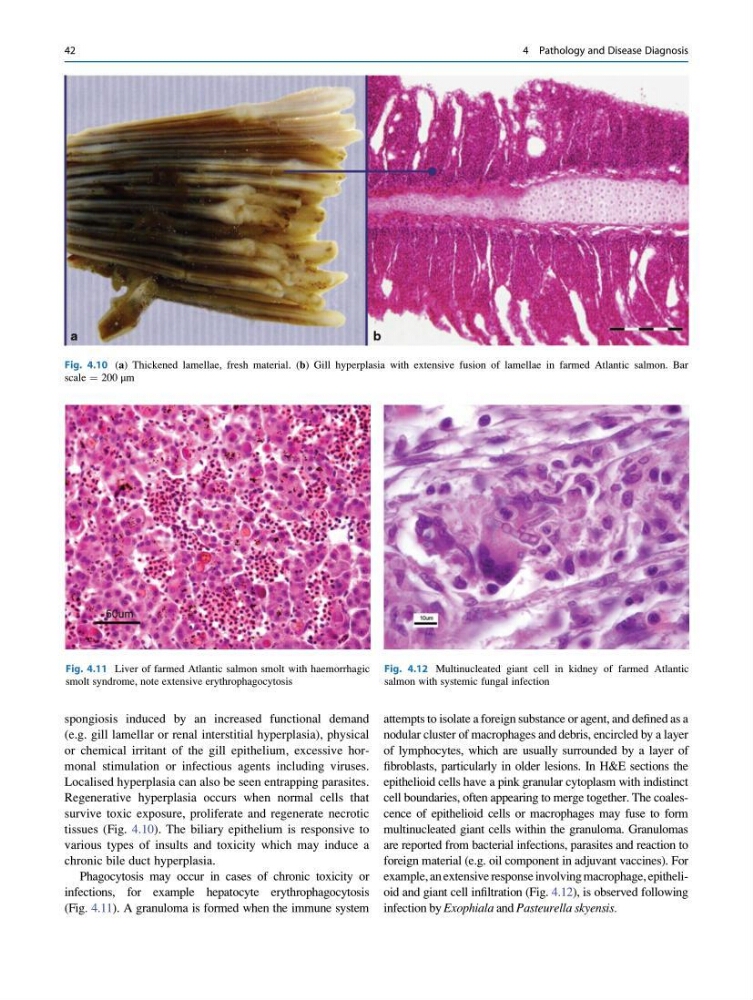
Fig. 4.10 (a) Thickened lamellae, fresh material. (b) Gill hyperplasia with extensive fusion of lamellae in farmed Atlantic salmon. Bar
scale ¼ 200 μm
Fig. 4.11 Liver of farmed Atlantic salmon smolt with haemorrhagic
smolt syndrome, note extensive erythrophagocytosis
Fig. 4.12 Multinucleated giant cell in kidney of farmed Atlantic
salmon with systemic fungal infection
42
4 Pathology and Disease Diagnosis
(e.g. gill lamellar or renal interstitial hyperplasia), physical
or chemical irritant of the gill epithelium, excessive hormonal stimulation or infectious agents including viruses.
Localised hyperplasia can also be seen entrapping parasites.
Regenerative hyperplasia occurs when normal cells that
survive toxic exposure, proliferate and regenerate necrotic
tissues (Fig. 4.10). The biliary epithelium is responsive to
various types of insults and toxicity which may induce a
chronic bile duct hyperplasia.
Phagocytosis may occur in cases of chronic toxicity or
infections, for example hepatocyte erythrophagocytosis
(Fig. 4.11). A granuloma is formed when the immune system
attempts to isolate a foreign substance or agent, and defined as a
nodular cluster of macrophages and debris, encircled by a layer
of lymphocytes, which are usually surrounded by a layer of
fibroblasts, particularly in older lesions. In H&E sections the
epithelioid cells have a pink granular cytoplasm with indistinct
cell boundaries, often appearing to merge together. The coalescence of epithelioid cells or macrophages may fuse to form
multinucleated giant cells within the granuloma. Granulomas
are reported from bacterial infections, parasites and reaction to
foreign material (e.g. oil component in adjuvant vaccines). For
example, an extensive response involving macrophage, epithelioid and giant cell infiltration (Fig. 4.12), is observed following
infection by Exophiala and Pasteurella skyensis.
Fig. 4.10 (a) Thickened lamellae, fresh material. (b) Gill hyperplasia with extensive fusion of lamellae in farmed Atlantic salmon. Bar
scale ¼ 200 μm
Fig. 4.11 Liver of farmed Atlantic salmon smolt with haemorrhagic
smolt syndrome, note extensive erythrophagocytosis
Fig. 4.12 Multinucleated giant cell in kidney of farmed Atlantic
salmon with systemic fungal infection
42
4 Pathology and Disease Diagnosis