

and cell sloughing. Damage to sub-surface cells show dramatic degeneration of the cytoplasm, although the nucleus
usually remains intact. In the recovery phase, large numbers
of eosinophilic granular cells may be seen within the
lamellae.
Morphometric data has proven that Ichthyobodo from
freshwater and sea water display a different cell shape
and data from electron microscopy show that marine
forms of Ichthyobodo possess ridge-like projections
along the cytostome process, which are smooth in parasites
from Atlantic salmon in fresh water. Phylogenetic analyses
of SSU rDNA sequences were able to prove the existence
of two Ichthyobodo species able to infect Atlantic salmon
based on differences in the attachment region and the
presence of spine-like surface projections, and the name
Ichthyobodo salmonis sp. n. has been proposed.
The diagnosis of Ichthyobodo is based on the microscopic examination of fresh tissues and the identification
of characteristic motile flagellates in mucous from the gills
or skin. Stained sections also demonstrate the attached
parasites.
8.5.3 Spironucleus spp.
Diplomonad flagellates are reported from several fish spices
worldwide and most of them are commensals feeding on
bacteria and on food digested by the host. However, some of
them are pathogenic and among salmonid species, they may
occur as enteric commensals or parasites (S. salmonis and
S. barkhanus), or they may cause severe systemic disease
(S. salmonicida). Intestinal diplomonads including infection
of the gallbladder occur commonly as opportunistic
parasites. In moderate numbers they seldom cause any
harm, but heavily infected fry and fingerlings, especially of
fresh water brook, brown, lake and rainbow trout, may show
nonspecific locomotive disorders, emaciation, catarrhal
enteritis, abdominal distension and exophthalmia. Gut
contents may be yellowish and fish produce a pseudo faeces.
Spironucleus salmonis, previously known as Hexamita
salmonis, had been known as a health issue during the
early life stages of the fresh water reared rainbow trout for
a long time, with high levels of morbidity and associated
mortality. Infections of the intestine of rainbow trout cause
weakness, anorexia and emaciation. Internally, enteritis,
intestinal haemorrhage, yellow mucus and necrosis of
hepatocytes may be observed.
S. barkhanus has been described from grayling and Arctic
char. In Northern Norway, systemic infection with
S. salmonicida has caused losses in several Atlantic salmon
sea farms. A large proportion of the large fish in the population
may be affected leading to rejects and downgrading. External
lesions may include ascites and exophthalmia, while internally, haemorrhagic boil-like lesions in the muscle and
necrotic patches in the kidney, spleen and liver, has been
reported for S. salmonicida and S. barkhanus (Figs. 8.26 and
8.27). Affected fish often have an unpleasant, putrid odour at
necropsy. Diffuse epicarditis or whitish cysts containing vast
numbers of parasites may be found on the ventricular wall
(Fig. 8.28). Microscopy also reveals widespread liquefactive
muscle necrosis with haemorrhage. Parasites may be present
in large numbers typically in the gut (Fig. 8.29) and
characterized by their pear-shape and paired anterior nuclei
(‘eyes’). Multifocal necrosis may also be found in kidney, liver
and spleen. The inflammatory response is variable depending
on temperature and age of lesions. Parasite aggregates may be
found in blood vessels and in the spongy myocardium. Purulent pericarditis with vast numbers of parasites and inflammatory cells may also occur (Fig. 8.30). S. salmonicida has also
caused systemic disease in sea-farmed Arctic char in northern
Norway, and in farmed Chinook salmon in BC, Canada.
In these cases, the parasites have been found in large numbers
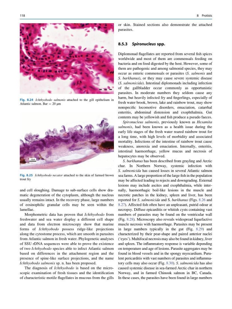
Fig. 8.24 Ichthyobodo salmonis attached to the gill epithelium in
Atlantic salmon. Bar ¼ 20 μm
Fig. 8.25 Ichthyobodo necator attached to the skin of farmed brown
trout fry
118
8 Protists
usually remains intact. In the recovery phase, large numbers
of eosinophilic granular cells may be seen within the
lamellae.
Morphometric data has proven that Ichthyobodo from
freshwater and sea water display a different cell shape
and data from electron microscopy show that marine
forms of Ichthyobodo possess ridge-like projections
along the cytostome process, which are smooth in parasites
from Atlantic salmon in fresh water. Phylogenetic analyses
of SSU rDNA sequences were able to prove the existence
of two Ichthyobodo species able to infect Atlantic salmon
based on differences in the attachment region and the
presence of spine-like surface projections, and the name
Ichthyobodo salmonis sp. n. has been proposed.
The diagnosis of Ichthyobodo is based on the microscopic examination of fresh tissues and the identification
of characteristic motile flagellates in mucous from the gills
or skin. Stained sections also demonstrate the attached
parasites.
8.5.3 Spironucleus spp.
Diplomonad flagellates are reported from several fish spices
worldwide and most of them are commensals feeding on
bacteria and on food digested by the host. However, some of
them are pathogenic and among salmonid species, they may
occur as enteric commensals or parasites (S. salmonis and
S. barkhanus), or they may cause severe systemic disease
(S. salmonicida). Intestinal diplomonads including infection
of the gallbladder occur commonly as opportunistic
parasites. In moderate numbers they seldom cause any
harm, but heavily infected fry and fingerlings, especially of
fresh water brook, brown, lake and rainbow trout, may show
nonspecific locomotive disorders, emaciation, catarrhal
enteritis, abdominal distension and exophthalmia. Gut
contents may be yellowish and fish produce a pseudo faeces.
Spironucleus salmonis, previously known as Hexamita
salmonis, had been known as a health issue during the
early life stages of the fresh water reared rainbow trout for
a long time, with high levels of morbidity and associated
mortality. Infections of the intestine of rainbow trout cause
weakness, anorexia and emaciation. Internally, enteritis,
intestinal haemorrhage, yellow mucus and necrosis of
hepatocytes may be observed.
S. barkhanus has been described from grayling and Arctic
char. In Northern Norway, systemic infection with
S. salmonicida has caused losses in several Atlantic salmon
sea farms. A large proportion of the large fish in the population
may be affected leading to rejects and downgrading. External
lesions may include ascites and exophthalmia, while internally, haemorrhagic boil-like lesions in the muscle and
necrotic patches in the kidney, spleen and liver, has been
reported for S. salmonicida and S. barkhanus (Figs. 8.26 and
8.27). Affected fish often have an unpleasant, putrid odour at
necropsy. Diffuse epicarditis or whitish cysts containing vast
numbers of parasites may be found on the ventricular wall
(Fig. 8.28). Microscopy also reveals widespread liquefactive
muscle necrosis with haemorrhage. Parasites may be present
in large numbers typically in the gut (Fig. 8.29) and
characterized by their pear-shape and paired anterior nuclei
(‘eyes’). Multifocal necrosis may also be found in kidney, liver
and spleen. The inflammatory response is variable depending
on temperature and age of lesions. Parasite aggregates may be
found in blood vessels and in the spongy myocardium. Purulent pericarditis with vast numbers of parasites and inflammatory cells may also occur (Fig. 8.30). S. salmonicida has also
caused systemic disease in sea-farmed Arctic char in northern
Norway, and in farmed Chinook salmon in BC, Canada.
In these cases, the parasites have been found in large numbers
Fig. 8.24 Ichthyobodo salmonis attached to the gill epithelium in
Atlantic salmon. Bar ¼ 20 μm
Fig. 8.25 Ichthyobodo necator attached to the skin of farmed brown
trout fry
118
8 Protists