

At necropsy, whitish nodules may be seen in many
organs, primarily the heart, muscle, kidney, liver and spleen.
Microscopically, a severe granulomatous response is characteristic, often with large numbers of macrophages and
multinucleated giant cells. Several developmental stages
may be observed, but a spore or resting stage in several
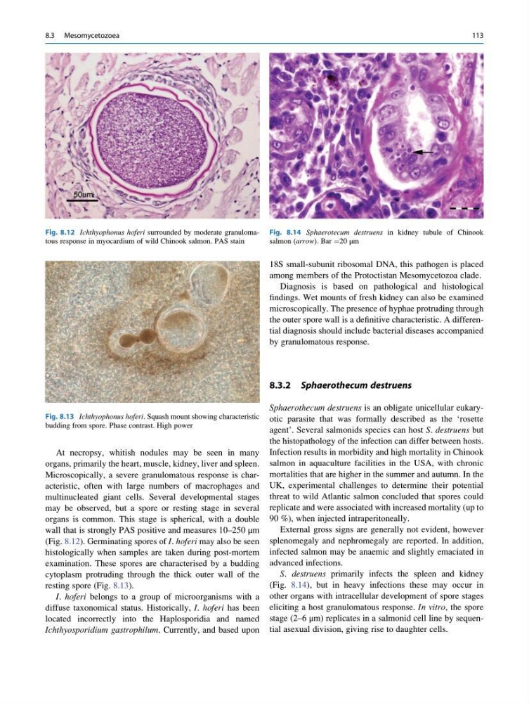
organs is common. This stage is spherical, with a double
wall that is strongly PAS positive and measures 10–250 μm
(Fig. 8.12). Germinating spores of I. hoferi may also be seen
histologically when samples are taken during post-mortem
examination. These spores are characterised by a budding
cytoplasm protruding through the thick outer wall of the
resting spore (Fig. 8.13).
I. hoferi belongs to a group of microorganisms with a
diffuse taxonomical status. Historically, I. hoferi has been
located incorrectly into the Haplosporidia and named
Ichthyosporidium gastrophilum. Currently, and based upon
18S small-subunit ribosomal DNA, this pathogen is placed
among members of the Protoctistan Mesomycetozoa clade.
Diagnosis is based on pathological and histological
findings. Wet mounts of fresh kidney can also be examined
microscopically. The presence of hyphae protruding through
the outer spore wall is a definitive characteristic. A differential diagnosis should include bacterial diseases accompanied
by granulomatous response.
8.3.2 Sphaerothecum destruens
Sphaerothecum destruens is an obligate unicellular eukaryotic parasite that was formally described as the ‘rosette
agent’. Several salmonids species can host S. destruens but
the histopathology of the infection can differ between hosts.
Infection results in morbidity and high mortality in Chinook
salmon in aquaculture facilities in the USA, with chronic
mortalities that are higher in the summer and autumn. In the
UK, experimental challenges to determine their potential
threat to wild Atlantic salmon concluded that spores could
replicate and were associated with increased mortality (up to
90 %), when injected intraperitoneally.
External gross signs are generally not evident, however
splenomegaly and nephromegaly are reported. In addition,
infected salmon may be anaemic and slightly emaciated in
advanced infections.
S. destruens primarily infects the spleen and kidney
(Fig. 8.14), but in heavy infections these may occur in
other organs with intracellular development of spore stages
eliciting a host granulomatous response. In vitro, the spore
stage (2–6 μm) replicates in a salmonid cell line by sequential asexual division, giving rise to daughter cells.
Fig. 8.12 Ichthyophonus hoferi surrounded by moderate granulomatous response in myocardium of wild Chinook salmon. PAS stain
Fig. 8.13 Ichthyophonus hoferi. Squash mount showing characteristic
budding from spore. Phase contrast. High power
Fig. 8.14 Sphaerotecum destruens in kidney tubule of Chinook
salmon (arrow). Bar ¼20 μm
8.3 Mesomycetozoea
113
organs, primarily the heart, muscle, kidney, liver and spleen.
Microscopically, a severe granulomatous response is characteristic, often with large numbers of macrophages and
multinucleated giant cells. Several developmental stages
may be observed, but a spore or resting stage in several
organs is common. This stage is spherical, with a double
wall that is strongly PAS positive and measures 10–250 μm
(Fig. 8.12). Germinating spores of I. hoferi may also be seen
histologically when samples are taken during post-mortem
examination. These spores are characterised by a budding
cytoplasm protruding through the thick outer wall of the
resting spore (Fig. 8.13).
I. hoferi belongs to a group of microorganisms with a
diffuse taxonomical status. Historically, I. hoferi has been
located incorrectly into the Haplosporidia and named
Ichthyosporidium gastrophilum. Currently, and based upon
18S small-subunit ribosomal DNA, this pathogen is placed
among members of the Protoctistan Mesomycetozoa clade.
Diagnosis is based on pathological and histological
findings. Wet mounts of fresh kidney can also be examined
microscopically. The presence of hyphae protruding through
the outer spore wall is a definitive characteristic. A differential diagnosis should include bacterial diseases accompanied
by granulomatous response.
8.3.2 Sphaerothecum destruens
Sphaerothecum destruens is an obligate unicellular eukaryotic parasite that was formally described as the ‘rosette
agent’. Several salmonids species can host S. destruens but
the histopathology of the infection can differ between hosts.
Infection results in morbidity and high mortality in Chinook
salmon in aquaculture facilities in the USA, with chronic
mortalities that are higher in the summer and autumn. In the
UK, experimental challenges to determine their potential
threat to wild Atlantic salmon concluded that spores could
replicate and were associated with increased mortality (up to
90 %), when injected intraperitoneally.
External gross signs are generally not evident, however
splenomegaly and nephromegaly are reported. In addition,
infected salmon may be anaemic and slightly emaciated in
advanced infections.
S. destruens primarily infects the spleen and kidney
(Fig. 8.14), but in heavy infections these may occur in
other organs with intracellular development of spore stages
eliciting a host granulomatous response. In vitro, the spore
stage (2–6 μm) replicates in a salmonid cell line by sequential asexual division, giving rise to daughter cells.
Fig. 8.12 Ichthyophonus hoferi surrounded by moderate granulomatous response in myocardium of wild Chinook salmon. PAS stain
Fig. 8.13 Ichthyophonus hoferi. Squash mount showing characteristic
budding from spore. Phase contrast. High power
Fig. 8.14 Sphaerotecum destruens in kidney tubule of Chinook
salmon (arrow). Bar ¼20 μm
8.3 Mesomycetozoea
113