

warmer temperatures, suspended organic matter, cage fouling,
high stocking densities and previous gill damage, have also
been reported as risk factors.
Primary attachment by amoeba is associated with branchial irritation and localized host cellular alterations including squamation-stratification of the epithelium (Fig. 8.2).
This is followed by epithelial hypertrophy and stratification
of epithelia at lesion surfaces, with recruitment of mucous
cells to affected areas, reduced chloride cells and the formation of large interlamellar lacunae or vesicles. Small to
medium sized lacunae may contain amoeba, whereas larger
lacunae are generally clear of any cellular debris (Fig. 8.3).
Very similar gill lesions associated with amoeba infections
on brook trout reared in freshwater, have also been reported
(Fig. 8.4). Proactive treatment by freshwater bathing is
carried out when gross gill assessment (i.e. a gill score)
indicates a moderate level of infestation in a population.
However, re-infections have been reported at 2 weeks post
bathing. During the post-bath period, non-AGD lesions
including haemorrhage, necrosis and regenerative hyperplasia have been observed.
Paramoeba can be detected from wet preparations of gill
tissue, although subsequent histological examination is
recommended to improve diagnostic accuracy. Histologically, hyperplasia, lamellar fusion, vesicles or lacunae, flattened epithelial cells (‘pavement cells’) and the presence of
amoeba form the basis of a diagnosis. Species identification
requires molecular testing using a quantitative duplex realtime PCR. Differential diagnosis includes other Protists,
water borne irritants and bacteria.
8.2
Ciliophora
8.2.1 Capriniana piscium
Capriniana (previously known as Trichophrya), is an
ectocommensal organism that commonly occurs on the
gills of several fresh water fish species. The shape of the
Table 8.1 Principal Protists described for salmonids
Name
Class or group
Common location
Environment
Paramoeba perurans
Amoebozoa
Gills
SW
Capriniana piscium
Ciliophora
Gills
FW
Chilodonella piscicola
Ciliophora
Gills, external surface
FW
Epistylis spp.
Ciliophora
Gills, external surface
FW
Ichthyophthirius multifiliis
Ciliophora
Oral cavity, gills, external surface
FW
Trichodina truttae
Ciliophora
Gills, external surface
FW, SW
Ichthyophonus hoferi
Mesomycetozoea
Musculature
FW, SW
Sphaerothecum destruens
Mesomycetozoea
Macrophages particularly in the spleen and kidney
FW
Dermocystidium salmonis
Mesomycetozoea
Oral cavity, external surface
FW, SW
Kabatana takedai
Microsporidia
Musculature, gills
FW, SW
Loma salmonae
Microsporidia
Gills
FW
Paranucleospora theridion
(Desmozoon lepeophtherii)
Microsporidia
Many organs
SW
Cryptobia salmositica
Sarcomastigophora
Blood
FW
Ichthyobodo spp.
Sarcomastigophora
Gills, external surface
FW, SW
Spironucleus salmonicida
Sarcomastigophora
Systemic
FW, SW
Spironucleus salmonis
Sarcomastigophora
Intestine
FW
Spironuclueus barkhanus
Sarcomastigophora
Gall bladder, intestine
FW
FW freshwater, SW sea water
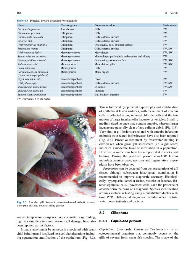
Fig. 8.1 Amoebic gill disease in seawater-farmed Atlantic salmon.
Note pale gills and nodular, slimy patches
108
8 Protists
high stocking densities and previous gill damage, have also
been reported as risk factors.
Primary attachment by amoeba is associated with branchial irritation and localized host cellular alterations including squamation-stratification of the epithelium (Fig. 8.2).
This is followed by epithelial hypertrophy and stratification
of epithelia at lesion surfaces, with recruitment of mucous
cells to affected areas, reduced chloride cells and the formation of large interlamellar lacunae or vesicles. Small to
medium sized lacunae may contain amoeba, whereas larger
lacunae are generally clear of any cellular debris (Fig. 8.3).
Very similar gill lesions associated with amoeba infections
on brook trout reared in freshwater, have also been reported
(Fig. 8.4). Proactive treatment by freshwater bathing is
carried out when gross gill assessment (i.e. a gill score)
indicates a moderate level of infestation in a population.
However, re-infections have been reported at 2 weeks post
bathing. During the post-bath period, non-AGD lesions
including haemorrhage, necrosis and regenerative hyperplasia have been observed.
Paramoeba can be detected from wet preparations of gill
tissue, although subsequent histological examination is
recommended to improve diagnostic accuracy. Histologically, hyperplasia, lamellar fusion, vesicles or lacunae, flattened epithelial cells (‘pavement cells’) and the presence of
amoeba form the basis of a diagnosis. Species identification
requires molecular testing using a quantitative duplex realtime PCR. Differential diagnosis includes other Protists,
water borne irritants and bacteria.
8.2
Ciliophora
8.2.1 Capriniana piscium
Capriniana (previously known as Trichophrya), is an
ectocommensal organism that commonly occurs on the
gills of several fresh water fish species. The shape of the
Table 8.1 Principal Protists described for salmonids
Name
Class or group
Common location
Environment
Paramoeba perurans
Amoebozoa
Gills
SW
Capriniana piscium
Ciliophora
Gills
FW
Chilodonella piscicola
Ciliophora
Gills, external surface
FW
Epistylis spp.
Ciliophora
Gills, external surface
FW
Ichthyophthirius multifiliis
Ciliophora
Oral cavity, gills, external surface
FW
Trichodina truttae
Ciliophora
Gills, external surface
FW, SW
Ichthyophonus hoferi
Mesomycetozoea
Musculature
FW, SW
Sphaerothecum destruens
Mesomycetozoea
Macrophages particularly in the spleen and kidney
FW
Dermocystidium salmonis
Mesomycetozoea
Oral cavity, external surface
FW, SW
Kabatana takedai
Microsporidia
Musculature, gills
FW, SW
Loma salmonae
Microsporidia
Gills
FW
Paranucleospora theridion
(Desmozoon lepeophtherii)
Microsporidia
Many organs
SW
Cryptobia salmositica
Sarcomastigophora
Blood
FW
Ichthyobodo spp.
Sarcomastigophora
Gills, external surface
FW, SW
Spironucleus salmonicida
Sarcomastigophora
Systemic
FW, SW
Spironucleus salmonis
Sarcomastigophora
Intestine
FW
Spironuclueus barkhanus
Sarcomastigophora
Gall bladder, intestine
FW
FW freshwater, SW sea water
Fig. 8.1 Amoebic gill disease in seawater-farmed Atlantic salmon.
Note pale gills and nodular, slimy patches
108
8 Protists