

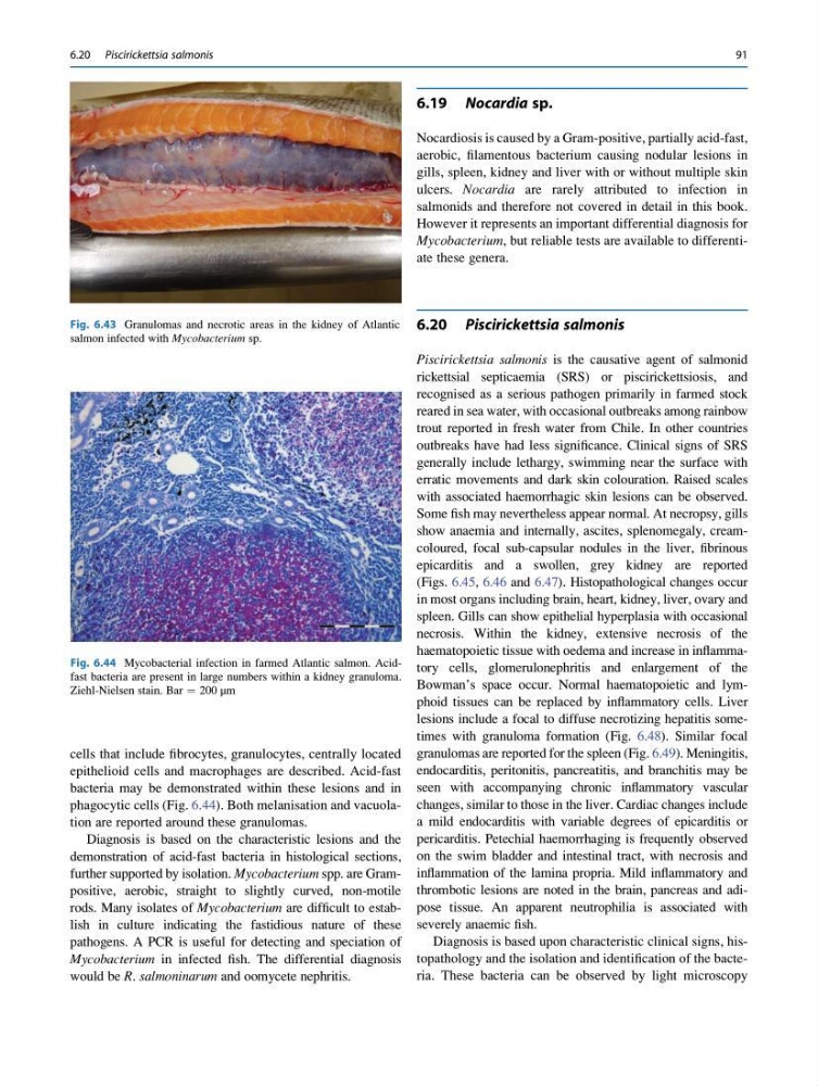
cells that include fibrocytes, granulocytes, centrally located
epithelioid cells and macrophages are described. Acid-fast
bacteria may be demonstrated within these lesions and in
phagocytic cells (Fig. 6.44). Both melanisation and vacuolation are reported around these granulomas.
Diagnosis is based on the characteristic lesions and the
demonstration of acid-fast bacteria in histological sections,
further supported by isolation. Mycobacterium spp. are Grampositive, aerobic, straight to slightly curved, non-motile
rods. Many isolates of Mycobacterium are difficult to establish in culture indicating the fastidious nature of these
pathogens. A PCR is useful for detecting and speciation of
Mycobacterium in infected fish. The differential diagnosis
would be R. salmoninarum and oomycete nephritis.
6.19 Nocardia sp.
Nocardiosis is caused by a Gram-positive, partially acid-fast,
aerobic, filamentous bacterium causing nodular lesions in
gills, spleen, kidney and liver with or without multiple skin
ulcers. Nocardia are rarely attributed to infection in
salmonids and therefore not covered in detail in this book.
However it represents an important differential diagnosis for
Mycobacterium, but reliable tests are available to differentiate these genera.
6.20 Piscirickettsia salmonis
Piscirickettsia salmonis is the causative agent of salmonid
rickettsial septicaemia (SRS) or piscirickettsiosis, and
recognised as a serious pathogen primarily in farmed stock
reared in sea water, with occasional outbreaks among rainbow
trout reported in fresh water from Chile. In other countries
outbreaks have had less significance. Clinical signs of SRS
generally include lethargy, swimming near the surface with
erratic movements and dark skin colouration. Raised scales
with associated haemorrhagic skin lesions can be observed.
Some fish may nevertheless appear normal. At necropsy, gills
show anaemia and internally, ascites, splenomegaly, creamcoloured, focal sub-capsular nodules in the liver, fibrinous
epicarditis and a swollen, grey kidney are reported
(Figs. 6.45, 6.46 and 6.47). Histopathological changes occur
in most organs including brain, heart, kidney, liver, ovary and
spleen. Gills can show epithelial hyperplasia with occasional
necrosis. Within the kidney, extensive necrosis of the
haematopoietic tissue with oedema and increase in inflammatory cells, glomerulonephritis and enlargement of the
Bowman’s space occur. Normal haematopoietic and lymphoid tissues can be replaced by inflammatory cells. Liver
lesions include a focal to diffuse necrotizing hepatitis sometimes with granuloma formation (Fig. 6.48). Similar focal
granulomas are reported for the spleen (Fig. 6.49). Meningitis,
endocarditis, peritonitis, pancreatitis, and branchitis may be
seen with accompanying chronic inflammatory vascular
changes, similar to those in the liver. Cardiac changes include
a mild endocarditis with variable degrees of epicarditis or
pericarditis. Petechial haemorrhaging is frequently observed
on the swim bladder and intestinal tract, with necrosis and
inflammation of the lamina propria. Mild inflammatory and
thrombotic lesions are noted in the brain, pancreas and adipose tissue. An apparent neutrophilia is associated with
severely anaemic fish.
Diagnosis is based upon characteristic clinical signs, histopathology and the isolation and identification of the bacteria. These bacteria can be observed by light microscopy
Fig. 6.43 Granulomas and necrotic areas in the kidney of Atlantic
salmon infected with Mycobacterium sp.
Fig. 6.44 Mycobacterial infection in farmed Atlantic salmon. Acidfast bacteria are present in large numbers within a kidney granuloma.
Ziehl-Nielsen stain. Bar ¼ 200 μm
6.20 Piscirickettsia salmonis
91
epithelioid cells and macrophages are described. Acid-fast
bacteria may be demonstrated within these lesions and in
phagocytic cells (Fig. 6.44). Both melanisation and vacuolation are reported around these granulomas.
Diagnosis is based on the characteristic lesions and the
demonstration of acid-fast bacteria in histological sections,
further supported by isolation. Mycobacterium spp. are Grampositive, aerobic, straight to slightly curved, non-motile
rods. Many isolates of Mycobacterium are difficult to establish in culture indicating the fastidious nature of these
pathogens. A PCR is useful for detecting and speciation of
Mycobacterium in infected fish. The differential diagnosis
would be R. salmoninarum and oomycete nephritis.
6.19 Nocardia sp.
Nocardiosis is caused by a Gram-positive, partially acid-fast,
aerobic, filamentous bacterium causing nodular lesions in
gills, spleen, kidney and liver with or without multiple skin
ulcers. Nocardia are rarely attributed to infection in
salmonids and therefore not covered in detail in this book.
However it represents an important differential diagnosis for
Mycobacterium, but reliable tests are available to differentiate these genera.
6.20 Piscirickettsia salmonis
Piscirickettsia salmonis is the causative agent of salmonid
rickettsial septicaemia (SRS) or piscirickettsiosis, and
recognised as a serious pathogen primarily in farmed stock
reared in sea water, with occasional outbreaks among rainbow
trout reported in fresh water from Chile. In other countries
outbreaks have had less significance. Clinical signs of SRS
generally include lethargy, swimming near the surface with
erratic movements and dark skin colouration. Raised scales
with associated haemorrhagic skin lesions can be observed.
Some fish may nevertheless appear normal. At necropsy, gills
show anaemia and internally, ascites, splenomegaly, creamcoloured, focal sub-capsular nodules in the liver, fibrinous
epicarditis and a swollen, grey kidney are reported
(Figs. 6.45, 6.46 and 6.47). Histopathological changes occur
in most organs including brain, heart, kidney, liver, ovary and
spleen. Gills can show epithelial hyperplasia with occasional
necrosis. Within the kidney, extensive necrosis of the
haematopoietic tissue with oedema and increase in inflammatory cells, glomerulonephritis and enlargement of the
Bowman’s space occur. Normal haematopoietic and lymphoid tissues can be replaced by inflammatory cells. Liver
lesions include a focal to diffuse necrotizing hepatitis sometimes with granuloma formation (Fig. 6.48). Similar focal
granulomas are reported for the spleen (Fig. 6.49). Meningitis,
endocarditis, peritonitis, pancreatitis, and branchitis may be
seen with accompanying chronic inflammatory vascular
changes, similar to those in the liver. Cardiac changes include
a mild endocarditis with variable degrees of epicarditis or
pericarditis. Petechial haemorrhaging is frequently observed
on the swim bladder and intestinal tract, with necrosis and
inflammation of the lamina propria. Mild inflammatory and
thrombotic lesions are noted in the brain, pancreas and adipose tissue. An apparent neutrophilia is associated with
severely anaemic fish.
Diagnosis is based upon characteristic clinical signs, histopathology and the isolation and identification of the bacteria. These bacteria can be observed by light microscopy
Fig. 6.43 Granulomas and necrotic areas in the kidney of Atlantic
salmon infected with Mycobacterium sp.
Fig. 6.44 Mycobacterial infection in farmed Atlantic salmon. Acidfast bacteria are present in large numbers within a kidney granuloma.
Ziehl-Nielsen stain. Bar ¼ 200 μm
6.20 Piscirickettsia salmonis
91