

reported. Retrograde extension from the posterior uvea to the
floor of the diencephalon along the epineurium and perineurium of the optic nerve, may also be a route of neural invasion.
Renibacterium salmoninarum occur within phagocytes and
readily multiply within making it possible to avoid the immune
mechanisms of the host and the effect of antibiotic therapy.
Diagnosis is based on the observation of typical clinical
signs and the histological observation of Gram positive bacilli
within tissues and phagocytes. R. salmoninarum can be
detected by Gram and PAS staining, but not by H&E.
R. salmoninarum is a small (0.5 Â 1.0 μm), slow-growing
Gram positive, non-acid fast, non-motile diplobacillus with
optimal growth at 15
C. The bacterium is proteolytic, produces
catalase and has an absolute requirement for L-cysteine.
Culture is achieved after several weeks on medium such as
Mueller Hinton with added L-cysteine hydrochloride. An
ELISA and a real time PCR are commonly used for diagnosis.
6.16 Carnobacterium maltaromaticum
Carnobacterium maltaromaticum (synonym C. piscicola)
infections are responsible of the condition recognised as ‘pseudo
kidney disease’. The bacterium has been be recovered from
rainbow trout, Chinook salmon and whitefish from North America and also, although less frequently, from fish within Europe,
Australia and South America. The genus Carnobacterium
incorporates Lactobacillus piscicola and related lactic acid
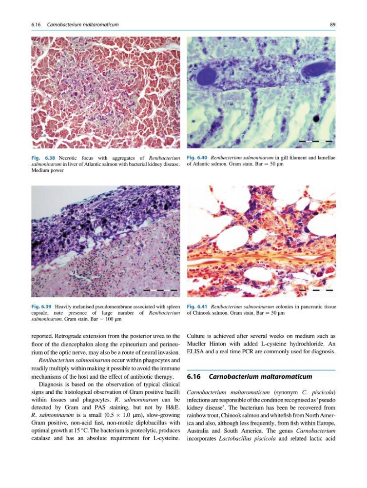
Fig. 6.38 Necrotic focus with aggregates of Renibacterium
salmoninarum in liver of Atlantic salmon with bacterial kidney disease.
Medium power
Fig. 6.39 Heavily melanised pseudomembrane associated with spleen
capsule, note presence of large number of Renibacterium
salmoninarum. Gram stain. Bar ¼ 100 μm
Fig. 6.40 Renibacterium salmoninarum in gill filament and lamellae
of Atlantic salmon. Gram stain. Bar ¼ 50 μm
Fig. 6.41 Renibacterium salmoninarum colonies in pancreatic tissue
of Chinook salmon. Gram stain. Bar ¼ 50 μm
6.16 Carnobacterium maltaromaticum
89
floor of the diencephalon along the epineurium and perineurium of the optic nerve, may also be a route of neural invasion.
Renibacterium salmoninarum occur within phagocytes and
readily multiply within making it possible to avoid the immune
mechanisms of the host and the effect of antibiotic therapy.
Diagnosis is based on the observation of typical clinical
signs and the histological observation of Gram positive bacilli
within tissues and phagocytes. R. salmoninarum can be
detected by Gram and PAS staining, but not by H&E.
R. salmoninarum is a small (0.5 Â 1.0 μm), slow-growing
Gram positive, non-acid fast, non-motile diplobacillus with
optimal growth at 15
C. The bacterium is proteolytic, produces
catalase and has an absolute requirement for L-cysteine.
Culture is achieved after several weeks on medium such as
Mueller Hinton with added L-cysteine hydrochloride. An
ELISA and a real time PCR are commonly used for diagnosis.
6.16 Carnobacterium maltaromaticum
Carnobacterium maltaromaticum (synonym C. piscicola)
infections are responsible of the condition recognised as ‘pseudo
kidney disease’. The bacterium has been be recovered from
rainbow trout, Chinook salmon and whitefish from North America and also, although less frequently, from fish within Europe,
Australia and South America. The genus Carnobacterium
incorporates Lactobacillus piscicola and related lactic acid
Fig. 6.38 Necrotic focus with aggregates of Renibacterium
salmoninarum in liver of Atlantic salmon with bacterial kidney disease.
Medium power
Fig. 6.39 Heavily melanised pseudomembrane associated with spleen
capsule, note presence of large number of Renibacterium
salmoninarum. Gram stain. Bar ¼ 100 μm
Fig. 6.40 Renibacterium salmoninarum in gill filament and lamellae
of Atlantic salmon. Gram stain. Bar ¼ 50 μm
Fig. 6.41 Renibacterium salmoninarum colonies in pancreatic tissue
of Chinook salmon. Gram stain. Bar ¼ 50 μm
6.16 Carnobacterium maltaromaticum
89