

31 Multicolor FISH Techniques for the Detection of Inter- and Intrachromosomal Rearrangements
419
~,
r- -~
1:1
•• .,
... "
,
~
.
1
?
"I
·1
l~
X
~'
II
"
,.~
2t .. ;
I •
••
Il
"'
II
'I
, n
11
1/
1.1
• ~
ttl
.. "
••
., ...
'.!
11
11
1',
It,
l'?
111
•• 1'I •• 1.0
. .
,:1
~, ,
y
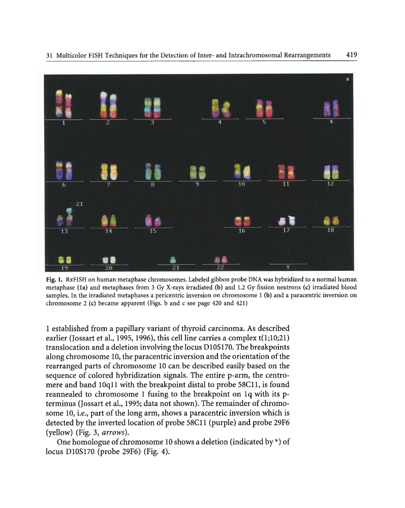
Fig. 1. RxFISH on human metaphase chromosomes. Labeled gibbon probe DNA was hybridized to a normal human
metaphase (la) and metaphases from 3 Gy X-rays irradiated (b) and 1.2 Gy fission neutrons (c) irradiated blood
samples. In the irradiated metaphases a pericentric inversion on chromosome 1 (b) and a paracentric inversion on
chromosome 2 (c) became apparent (Figs. band c see page 420 and 421)
1 established from a papillary variant of thyroid carcinoma. As described
earlier (Jossart et al., 1995, 1996), this cell line carries a complex t(1;10;21)
translocation and a deletion involving the locus DI0S170. The breakpoints
along chromosome 10, the paracentric inversion and the orientation of the
rearranged parts of chromosome 10 can be described easily based on the
sequence of colored hybridization signals. The entire p-arm, the centromere and band lOqll with the breakpoint distal to probe S8Cll, is found
reannealed to chromosome 1 fusing to the breakpoint on lq with its pterminus (Jossart et aI., 1995; data not shown). The remainder of chromosome 10, i.e., part of the long arm, shows a paracentric inversion which is
detected by the inverted location of probe S8Cli (purple) and probe 29F6
(yellow) (Fig. 3, arrows).
One homologue of chromosome 10 shows a deletion (indicated by *) of
locus DI0S170 (probe 29F6) (Fig. 4).
419
~,
r- -~
1:1
•• .,
... "
,
~
.
1
?
"I
·1
l~
X
~'
II
"
,.~
2t .. ;
I •
••
Il
"'
II
'I
, n
11
1/
1.1
• ~
ttl
.. "
••
., ...
'.!
11
11
1',
It,
l'?
111
•• 1'I •• 1.0
. .
,:1
~, ,
y
Fig. 1. RxFISH on human metaphase chromosomes. Labeled gibbon probe DNA was hybridized to a normal human
metaphase (la) and metaphases from 3 Gy X-rays irradiated (b) and 1.2 Gy fission neutrons (c) irradiated blood
samples. In the irradiated metaphases a pericentric inversion on chromosome 1 (b) and a paracentric inversion on
chromosome 2 (c) became apparent (Figs. band c see page 420 and 421)
1 established from a papillary variant of thyroid carcinoma. As described
earlier (Jossart et al., 1995, 1996), this cell line carries a complex t(1;10;21)
translocation and a deletion involving the locus DI0S170. The breakpoints
along chromosome 10, the paracentric inversion and the orientation of the
rearranged parts of chromosome 10 can be described easily based on the
sequence of colored hybridization signals. The entire p-arm, the centromere and band lOqll with the breakpoint distal to probe S8Cll, is found
reannealed to chromosome 1 fusing to the breakpoint on lq with its pterminus (Jossart et aI., 1995; data not shown). The remainder of chromosome 10, i.e., part of the long arm, shows a paracentric inversion which is
detected by the inverted location of probe S8Cli (purple) and probe 29F6
(yellow) (Fig. 3, arrows).
One homologue of chromosome 10 shows a deletion (indicated by *) of
locus DI0S170 (probe 29F6) (Fig. 4).