

402
ILSE CHUDOBA and GABRIELE SENGER
somes from prenatal cases are analyzed, time is the most limiting factor
and repetition of microdissection experiments has to be avoided. We recommend collecting fragments into two different independent PCR tubes.
In principle one single fragment or chromosome is enough to obtain a
sufficiently strong fluorescence signal after hybridization. However, to
be on the safe side, we recommend taking 3 fragments or chromosomes
for each PCR tube.
After labeling of the PCR products, the probes should be hybridized to
normal metaphase spreads as well as back to metaphases from the patient.
This provides a good control for the correctness of the microdissection.
However, reverse FISH results can be confusing. One has to be aware that
the microdissection library hybridizes not only to the aberrant chromosome of the patient's metaphases, but to the corresponding normal chromosomes and chromosomal regions that are involved in the aberrant
chromosome. An example is given in Fig. 2. A derivative chromosome
21 was found prenatally in the case of a girl with severe dysmorphic features and developmental delay (Novotna et al. 1998). Microdissection of
the whole chromosome revealed the origin of the additional material as
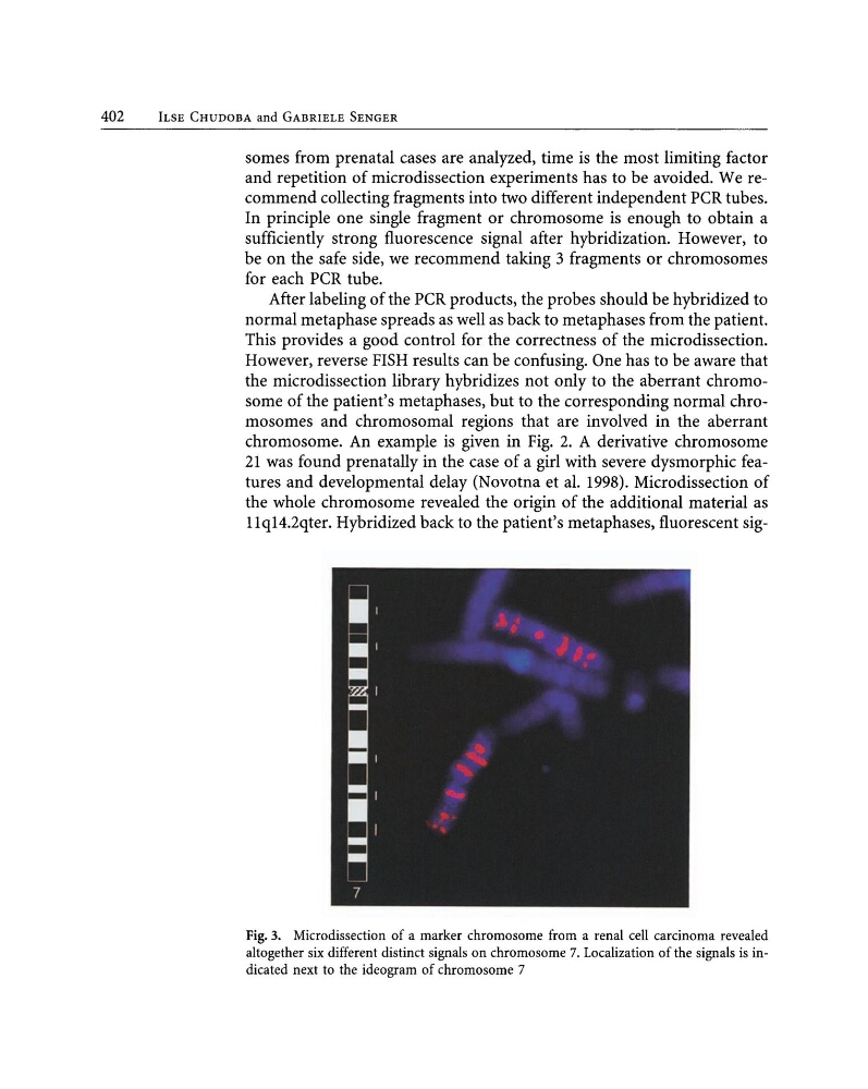
llqI4.2qter. Hybridized back to the patient's metaphases, fluorescent sigFig. 3. Microdissection of a marker chromosome from a renal cell carcinoma revealed
altogether six different distinct signals on chromosome 7. Localization of the signals is indicated next to the ideogram of chromosome 7
ILSE CHUDOBA and GABRIELE SENGER
somes from prenatal cases are analyzed, time is the most limiting factor
and repetition of microdissection experiments has to be avoided. We recommend collecting fragments into two different independent PCR tubes.
In principle one single fragment or chromosome is enough to obtain a
sufficiently strong fluorescence signal after hybridization. However, to
be on the safe side, we recommend taking 3 fragments or chromosomes
for each PCR tube.
After labeling of the PCR products, the probes should be hybridized to
normal metaphase spreads as well as back to metaphases from the patient.
This provides a good control for the correctness of the microdissection.
However, reverse FISH results can be confusing. One has to be aware that
the microdissection library hybridizes not only to the aberrant chromosome of the patient's metaphases, but to the corresponding normal chromosomes and chromosomal regions that are involved in the aberrant
chromosome. An example is given in Fig. 2. A derivative chromosome
21 was found prenatally in the case of a girl with severe dysmorphic features and developmental delay (Novotna et al. 1998). Microdissection of
the whole chromosome revealed the origin of the additional material as
llqI4.2qter. Hybridized back to the patient's metaphases, fluorescent sigFig. 3. Microdissection of a marker chromosome from a renal cell carcinoma revealed
altogether six different distinct signals on chromosome 7. Localization of the signals is indicated next to the ideogram of chromosome 7