

394
ILSE CHUDOBA and GABRIELE SENGER
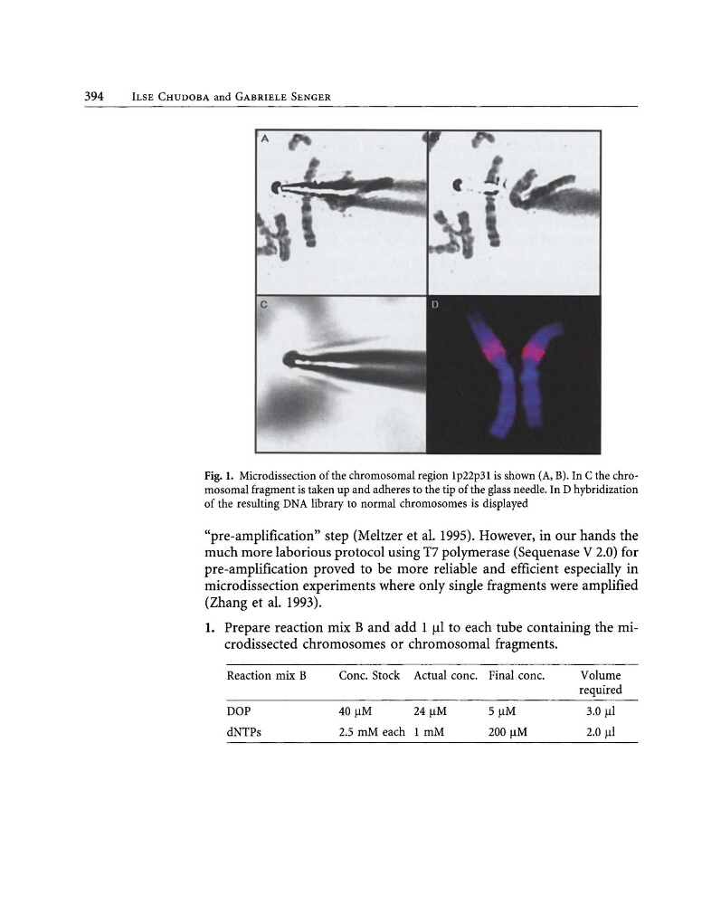
Fig. 1. Microdissection of the chromosomal region Ip22p3l is shown (A, B). In C the chromosomal fragment is taken up and adheres to the tip of the glass needle. In D hybridization
of the resulting DNA library to normal chromosomes is displayed
"pre-amplification" step (Meltzer et al. 1995). However, in our hands the
much more laborious protocol using T7 polymerase (Sequenase V 2.0) for
pre-amplification proved to be more reliable and efficient especially in
microdissection experiments where only single fragments were amplified
(Zhang et al. 1993).
1. Prepare reaction mix B and add 1 III to each tube containing the microdissected chromosomes or chromosomal fragments.
Reaction mix B
Cone. Stock Actual conc. Final conc.
Volume
required
DOP
40 11M
24 11M
5 11M
3.0 III
dNTPs
2.5 mM each I mM
200 11M
2.0 III
ILSE CHUDOBA and GABRIELE SENGER
Fig. 1. Microdissection of the chromosomal region Ip22p3l is shown (A, B). In C the chromosomal fragment is taken up and adheres to the tip of the glass needle. In D hybridization
of the resulting DNA library to normal chromosomes is displayed
"pre-amplification" step (Meltzer et al. 1995). However, in our hands the
much more laborious protocol using T7 polymerase (Sequenase V 2.0) for
pre-amplification proved to be more reliable and efficient especially in
microdissection experiments where only single fragments were amplified
(Zhang et al. 1993).
1. Prepare reaction mix B and add 1 III to each tube containing the microdissected chromosomes or chromosomal fragments.
Reaction mix B
Cone. Stock Actual conc. Final conc.
Volume
required
DOP
40 11M
24 11M
5 11M
3.0 III
dNTPs
2.5 mM each I mM
200 11M
2.0 III