

324
THOMAS LIEHR
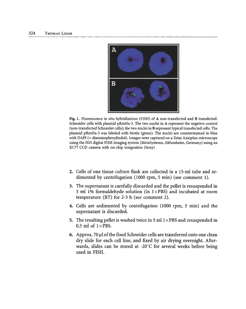
Fig. 1. Fluorescence in situ hybridization (FISH) of A non-transfected and B transfected.
Schneider cells with plasmid pRmHa-3. The two nuclei in A represent the negative control
(non-transfected Schneider cells); the two nuclei in B represent typical transfected cells. The
plasmid pRmHa-3 was labeled with biotin (green). The nuclei are counterstained in blue
with DAPI (= diaminophenylindol). Images were captured on a Zeiss Axioplan microscope
using the ISIS digital FISH imaging system (MetaSystems, Altlussheim, Germany) using an
XC77 CCD camera with on-chip integration (Sony)
2. Cells of one tissue culture flask are collected in a 15-ml tube and sedimented by centrifugation (1000 rpm,S min) (see comment 1).
3. The supernatant is carefully discarded and the pellet is resuspended in
5 ml I% formaldehyde solution (in I x PBS) and incubated at room
temperature (RT) for 2-3 h (see comment 2).
4. Cells are sedimented by centrifugation (1000 rpm,S min) and the
supernatant is discarded.
5. The resulting pellet is washed twice in 5 mIl x PBS and resuspended in
0.5 ml of I x PBS.
6. Approx. 70 ....1of the ftxed Schneider cells are transferred onto one clean
dry slide for each cell line, and ftxed by air drying overnight. Afterwards, slides can be stored at -20°C for several weeks before being
used in FISH.
THOMAS LIEHR
Fig. 1. Fluorescence in situ hybridization (FISH) of A non-transfected and B transfected.
Schneider cells with plasmid pRmHa-3. The two nuclei in A represent the negative control
(non-transfected Schneider cells); the two nuclei in B represent typical transfected cells. The
plasmid pRmHa-3 was labeled with biotin (green). The nuclei are counterstained in blue
with DAPI (= diaminophenylindol). Images were captured on a Zeiss Axioplan microscope
using the ISIS digital FISH imaging system (MetaSystems, Altlussheim, Germany) using an
XC77 CCD camera with on-chip integration (Sony)
2. Cells of one tissue culture flask are collected in a 15-ml tube and sedimented by centrifugation (1000 rpm,S min) (see comment 1).
3. The supernatant is carefully discarded and the pellet is resuspended in
5 ml I% formaldehyde solution (in I x PBS) and incubated at room
temperature (RT) for 2-3 h (see comment 2).
4. Cells are sedimented by centrifugation (1000 rpm,S min) and the
supernatant is discarded.
5. The resulting pellet is washed twice in 5 mIl x PBS and resuspended in
0.5 ml of I x PBS.
6. Approx. 70 ....1of the ftxed Schneider cells are transferred onto one clean
dry slide for each cell line, and ftxed by air drying overnight. Afterwards, slides can be stored at -20°C for several weeks before being
used in FISH.