

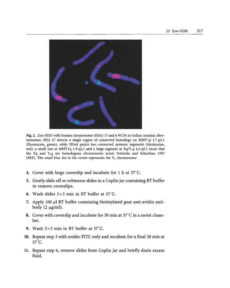
Fig. 2. Zoo-FISH with human chromosome (HSA) 17 and 4 WCPs to Indian muntjac chromosomes. HSA 17 detects a single region of conserved homology on MMVIp 1.7-p2.I
(fluorescein, green), while HSA4 paints two conserved syntenic segments (rhodamine,
red): a small one at MMVIq 1.9-q2.I and a large segment at Xq/Y1q 4.2-q5.I (note that
the Xq and Y1q are homologous chromosome arms) Fronicke and Scherthan 1997
(REF). The small blue dot in the center represents the Y2 chromosome
4. Cover with large coverslip and incubate for 1 h at 37°C.
5. Gently slide off or submerse slides in a Coplin jar containing BT buffer
to remove coverslips.
6. Wash slides 3x3 min in BT buffer at 37()C.
7. Apply 100 III BT buffer containing biotinylated goat anti-avidin antibody (2 Ilg/ml).
8. Cover with coverslip and incubate for 30 min at 37°C in a moist chamber.
9. Wash 3x3 min in BT buffer at 37°C.
10. Repeat step 3 with avidin-FITC only and incubate for a final 30 min at
37°C.
11. Repeat step 6, remove slides from Coplin jar and briefly drain excess
fluid.
25 Zoo-FISH
317
(fluorescein, green), while HSA4 paints two conserved syntenic segments (rhodamine,
red): a small one at MMVIq 1.9-q2.I and a large segment at Xq/Y1q 4.2-q5.I (note that
the Xq and Y1q are homologous chromosome arms) Fronicke and Scherthan 1997
(REF). The small blue dot in the center represents the Y2 chromosome
4. Cover with large coverslip and incubate for 1 h at 37°C.
5. Gently slide off or submerse slides in a Coplin jar containing BT buffer
to remove coverslips.
6. Wash slides 3x3 min in BT buffer at 37()C.
7. Apply 100 III BT buffer containing biotinylated goat anti-avidin antibody (2 Ilg/ml).
8. Cover with coverslip and incubate for 30 min at 37°C in a moist chamber.
9. Wash 3x3 min in BT buffer at 37°C.
10. Repeat step 3 with avidin-FITC only and incubate for a final 30 min at
37°C.
11. Repeat step 6, remove slides from Coplin jar and briefly drain excess
fluid.
25 Zoo-FISH
317