

278
JAIME GOSA.LVEZ et al.
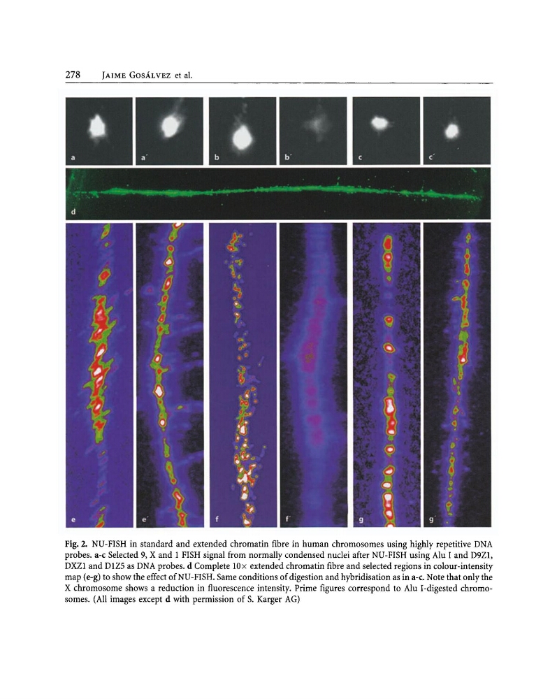
Fig. 2. NU-FISH in standard and extended chromatin fibre in human chromosomes using higWy repetitive DNA
probes. a-c Selected 9, X and I FISH signal from normally condensed nuclei after NU-FISH using Alu I and D9ZI,
DXZI and DIZS as DNA probes. d Complete lOx extended chromatin fibre and selected regions in colour-intensity
map (e-g) to show the effect ofNU-FISH. Same conditions of digestion and hybridisation as in a-c. Note that only the
X chromosome shows a reduction in fluorescence intensity. Prime figures correspond to Alu I-digested chromosomes. (All images except d with permission of S. Karger AG)
JAIME GOSA.LVEZ et al.
Fig. 2. NU-FISH in standard and extended chromatin fibre in human chromosomes using higWy repetitive DNA
probes. a-c Selected 9, X and I FISH signal from normally condensed nuclei after NU-FISH using Alu I and D9ZI,
DXZI and DIZS as DNA probes. d Complete lOx extended chromatin fibre and selected regions in colour-intensity
map (e-g) to show the effect ofNU-FISH. Same conditions of digestion and hybridisation as in a-c. Note that only the
X chromosome shows a reduction in fluorescence intensity. Prime figures correspond to Alu I-digested chromosomes. (All images except d with permission of S. Karger AG)