

222
BERND RAUTENSTRAUSS and CHRISTINA FUCHS
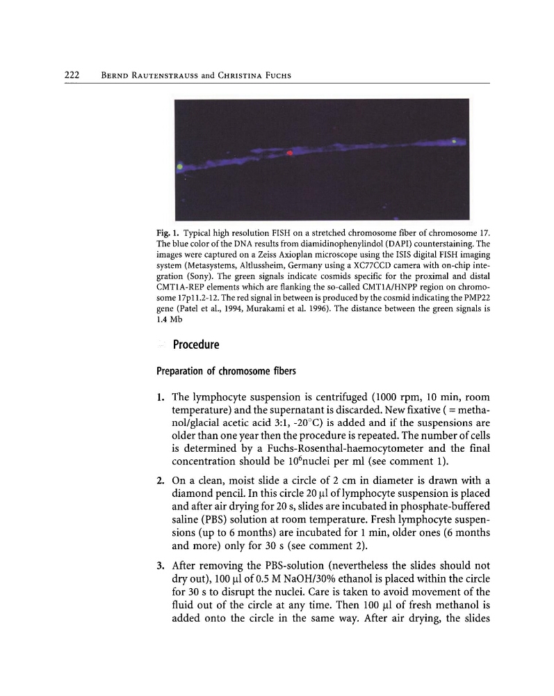
Fig. 1. Typical high resolution FISH on a stretched chromosome fiber of chromosome 17.
The blue color of the DNA results from diamidinophenylindol (DAPI) counterstaining. The
images were captured on a Zeiss Axioplan microscope using the ISIS digital FISH imaging
system (Metasystems, Altlussheim, Germany using a XC77CCD camera with on-chip integration (Sony). The green signals indicate cosmids specific for the proximal and distal
CMTlA-REP elements which are flanking the so-called CMTlA/HNPP region on chromosome 17p11.2-12. The red signal in between is produced by the cosmid indicating the PMP22
gene (Patel et aI., 1994, Murakami et al. 1996). The distance between the green signals is
1.4 Mb
Procedure
Preparation of chromosome fibers
1. The lymphocyte suspension is centrifuged (1000 rpm, 10 min, room
temperature) and the supernatant is discarded. New fIxative ( =methanol/glacial acetic acid 3:1, -20°C) is added and if the suspensions are
older than one year then the procedure is repeated. The number of cells
is determined by a Fuchs-Rosenthal-haemocytometer and the fInal
concentration should be 10
6
nuclei per ml (see comment 1).
2. On a clean, moist slide a circle of 2 cm in diameter is drawn with a
diamond pencil. In this circle 20 f.ll oflymphocyte suspension is placed
and after air drying for 20 s, slides are incubated in phosphate-buffered
saline (PBS) solution at room temperature. Fresh lymphocyte suspensions (up to 6 months) are incubated for 1 min, older ones (6 months
and more) only for 30 s (see comment 2).
3. After removing the PBS-solution (nevertheless the slides should not
dry out), 100 f.ll of 0.5 M NaOH/300/0 ethanol is placed within the circle
for 30 s to disrupt the nuclei. Care is taken to avoid movement of the
fluid out of the circle at any time. Then 100 f.ll of fresh methanol is
added onto the circle in the same way. After air drying, the slides
BERND RAUTENSTRAUSS and CHRISTINA FUCHS
Fig. 1. Typical high resolution FISH on a stretched chromosome fiber of chromosome 17.
The blue color of the DNA results from diamidinophenylindol (DAPI) counterstaining. The
images were captured on a Zeiss Axioplan microscope using the ISIS digital FISH imaging
system (Metasystems, Altlussheim, Germany using a XC77CCD camera with on-chip integration (Sony). The green signals indicate cosmids specific for the proximal and distal
CMTlA-REP elements which are flanking the so-called CMTlA/HNPP region on chromosome 17p11.2-12. The red signal in between is produced by the cosmid indicating the PMP22
gene (Patel et aI., 1994, Murakami et al. 1996). The distance between the green signals is
1.4 Mb
Procedure
Preparation of chromosome fibers
1. The lymphocyte suspension is centrifuged (1000 rpm, 10 min, room
temperature) and the supernatant is discarded. New fIxative ( =methanol/glacial acetic acid 3:1, -20°C) is added and if the suspensions are
older than one year then the procedure is repeated. The number of cells
is determined by a Fuchs-Rosenthal-haemocytometer and the fInal
concentration should be 10
6
nuclei per ml (see comment 1).
2. On a clean, moist slide a circle of 2 cm in diameter is drawn with a
diamond pencil. In this circle 20 f.ll oflymphocyte suspension is placed
and after air drying for 20 s, slides are incubated in phosphate-buffered
saline (PBS) solution at room temperature. Fresh lymphocyte suspensions (up to 6 months) are incubated for 1 min, older ones (6 months
and more) only for 30 s (see comment 2).
3. After removing the PBS-solution (nevertheless the slides should not
dry out), 100 f.ll of 0.5 M NaOH/300/0 ethanol is placed within the circle
for 30 s to disrupt the nuclei. Care is taken to avoid movement of the
fluid out of the circle at any time. Then 100 f.ll of fresh methanol is
added onto the circle in the same way. After air drying, the slides