

15 Preparation of Samples for CGH by DOP-PCR from microdissected cells
193
3. Remove tubes from PCR machine and add the following components
to each tube:
- 5 III Taq-buffer
- 5 III dNTP-mix (2 IlM)
- 5 III DOP-primer (10 IlM)
- 1 III Taq polymerase (5 units)
4. Fill with ultrapure water to a volume of 50 Ill.
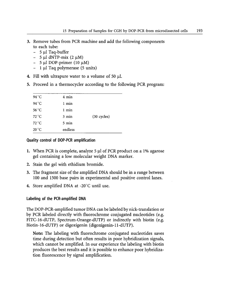
5. Proceed in a thermocyc1er according to the following PCR program:
94°C
4 min
94°C
1 min
56"C
1 min
12°C
3 min
(30 cycles)
12 °C
5 min
20°C
endless
Quality control of DOP-PCR amplification
1. When PCR is complete, analyze 5 III of PCR product on a 1% agarose
gel containing a low molecular weight DNA marker.
2. Stain the gel with ethidium bromide.
3. The fragment size of the amplified DNA should be in a range between
100 and 1500 base pairs in experimental and positive control lanes.
4. Store amplified DNA at -20°C until use.
labeling of the PCR-amplified DNA
The DOP-PCR-amplified tumor DNA can be labeled by nick-translation or
by PCR labeled directly with fluorochrome conjugated nuc1eotides (e.g.
FITC-16-dUTP, Spectrum-Orange-dUTP) or indirectly with biotin (e.g.
Biotin-16-dUTP) or digoxigenin (digoxigenin-ll-dUTP).
Note: The labeling with fluorochrome conjugated nuc1eotides saves
time during detection but often results in poor hybridization signals,
which cannot be amplified. In our experience the labeling with biotin
produces the best results and it is possible to enhance poor hybridization fluorescence by signal amplification.
193
3. Remove tubes from PCR machine and add the following components
to each tube:
- 5 III Taq-buffer
- 5 III dNTP-mix (2 IlM)
- 5 III DOP-primer (10 IlM)
- 1 III Taq polymerase (5 units)
4. Fill with ultrapure water to a volume of 50 Ill.
5. Proceed in a thermocyc1er according to the following PCR program:
94°C
4 min
94°C
1 min
56"C
1 min
12°C
3 min
(30 cycles)
12 °C
5 min
20°C
endless
Quality control of DOP-PCR amplification
1. When PCR is complete, analyze 5 III of PCR product on a 1% agarose
gel containing a low molecular weight DNA marker.
2. Stain the gel with ethidium bromide.
3. The fragment size of the amplified DNA should be in a range between
100 and 1500 base pairs in experimental and positive control lanes.
4. Store amplified DNA at -20°C until use.
labeling of the PCR-amplified DNA
The DOP-PCR-amplified tumor DNA can be labeled by nick-translation or
by PCR labeled directly with fluorochrome conjugated nuc1eotides (e.g.
FITC-16-dUTP, Spectrum-Orange-dUTP) or indirectly with biotin (e.g.
Biotin-16-dUTP) or digoxigenin (digoxigenin-ll-dUTP).
Note: The labeling with fluorochrome conjugated nuc1eotides saves
time during detection but often results in poor hybridization signals,
which cannot be amplified. In our experience the labeling with biotin
produces the best results and it is possible to enhance poor hybridization fluorescence by signal amplification.