

15 Preparation of Samples for CGH by DOP-PCR from microdissected cells
187
Outline
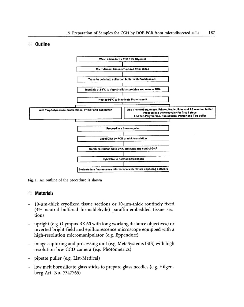
Wash .lId•• ln 1 x PBS fl',!, Glycerol
Microdls••cttis.u. structure. from slide.
Transfer cell. Into collection buffer with Protelnas.-K
Incubate at 55·C to dlg••t c.llular prot.lns .nd r.I•••• DNA
Heat to 95·C to In.ctivat. Protelnase-K
Add Taq-Polym.rase, Nucleotld••, Primer .nd T.q·buff.r
Add Th.rmoSequena... Primer, Nucleotldes .nd TS ...cUon buff.r
Proc••d In a thermocycl.r for flrst 8 .t.p.
Add T.q-Polymer•••• NucleoUdaa. Primer .nd T.q-buff.r
Combin. Hum.n Cot1-DNA. test-DNA and control-llNA
Hybridize to normal m.taphas••
Evaluat. in a fluor••c.nc. mlcroscop. with plctur. capturing .oftwar.
Fig. 1. An outline of the procedure is shown
' ," Materials
lO-l!m-thick cryofixed tissue sections or lO-l!m-thick routinely fixed
(4% neutral buffered formaldehyde) paraffin-embedded tissue sections
upright (e.g. Olympus BX 60 with long working distance objectives) or
inverted bright-field and epifluorescence microscope equipped with a
high-resolution micromanipulator (e.g. Eppendorf)
image capturing and processing unit (e.g. MetaSystems ISIS) with high
resolution b/w CCD camera (e.g. Photometrics)
pipette puller (e.g. List-Medical)
low melt borosilicate glass sticks to prepare glass needles (e.g. Hilgenberg Art. No. 7347765)
187
Outline
Wash .lId•• ln 1 x PBS fl',!, Glycerol
Microdls••cttis.u. structure. from slide.
Transfer cell. Into collection buffer with Protelnas.-K
Incubate at 55·C to dlg••t c.llular prot.lns .nd r.I•••• DNA
Heat to 95·C to In.ctivat. Protelnase-K
Add Taq-Polym.rase, Nucleotld••, Primer .nd T.q·buff.r
Add Th.rmoSequena... Primer, Nucleotldes .nd TS ...cUon buff.r
Proc••d In a thermocycl.r for flrst 8 .t.p.
Add T.q-Polymer•••• NucleoUdaa. Primer .nd T.q-buff.r
Combin. Hum.n Cot1-DNA. test-DNA and control-llNA
Hybridize to normal m.taphas••
Evaluat. in a fluor••c.nc. mlcroscop. with plctur. capturing .oftwar.
Fig. 1. An outline of the procedure is shown
' ," Materials
lO-l!m-thick cryofixed tissue sections or lO-l!m-thick routinely fixed
(4% neutral buffered formaldehyde) paraffin-embedded tissue sections
upright (e.g. Olympus BX 60 with long working distance objectives) or
inverted bright-field and epifluorescence microscope equipped with a
high-resolution micromanipulator (e.g. Eppendorf)
image capturing and processing unit (e.g. MetaSystems ISIS) with high
resolution b/w CCD camera (e.g. Photometrics)
pipette puller (e.g. List-Medical)
low melt borosilicate glass sticks to prepare glass needles (e.g. Hilgenberg Art. No. 7347765)