

10
EIGIL KJELDSEN and STEEN K0LVRAA
Once cells or tissues have been fIxed, the nucleic acids contained within
them have to be made available to the probe (unmasking). This process
often involves a proteolytic step most frequently using proteinase K or
pepsin Hel although other enzymes have been used. This step removes
components of the cell nucleus and cytoplasm to allow probe access.
Thus, under-digestion leads to suboptimal probe penetration whereas
over-digestion leads to destruction of chromosome, cell and tissue structure resulting in the loss of both nucleic acids and morphological detail.
When archival wax embedded tissues are used, a dewaxing step is also
required. Following proteolysis post-fIxation, particularly in aldehydebased fIxatives (e.g. paraformaldehyde), seems useful for the prevention
of further loss of material from the slide.
Ad 2: Probe source,
labeling and
preparation
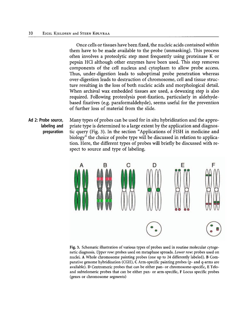
Many types of probes can be used for in situ hybridization and the appropriate type is determined to a large extent by the application and diagnostic query (Fig. 3). In the section "Applications of FISH in medicine and
biology" the choice of probe type will be discussed in relation to application. Here, the different types of probes will briefly be discussed with respect to source and type of labeling.
A
B
c
o
E
F
Fig. 3. Schematic illustration of various types of probes used in routine molecular cytogenetic diagnosis. Upper row: probes used on metaphase spreads. Lower row: probes used on
nuclei. A Whole chromosome painting probes (one up to 24 differently labeled). B Comparative genome hybridization (CGH), C Arm-specific painting probes (p- and q-arms are
available). D Centromeric probes that can be either pan- or chromosome-specific, E Teloand subtelomeric probes that can be either pan- or arm-specific, F Locus specific probes
(genes or chromosome segments)
EIGIL KJELDSEN and STEEN K0LVRAA
Once cells or tissues have been fIxed, the nucleic acids contained within
them have to be made available to the probe (unmasking). This process
often involves a proteolytic step most frequently using proteinase K or
pepsin Hel although other enzymes have been used. This step removes
components of the cell nucleus and cytoplasm to allow probe access.
Thus, under-digestion leads to suboptimal probe penetration whereas
over-digestion leads to destruction of chromosome, cell and tissue structure resulting in the loss of both nucleic acids and morphological detail.
When archival wax embedded tissues are used, a dewaxing step is also
required. Following proteolysis post-fIxation, particularly in aldehydebased fIxatives (e.g. paraformaldehyde), seems useful for the prevention
of further loss of material from the slide.
Ad 2: Probe source,
labeling and
preparation
Many types of probes can be used for in situ hybridization and the appropriate type is determined to a large extent by the application and diagnostic query (Fig. 3). In the section "Applications of FISH in medicine and
biology" the choice of probe type will be discussed in relation to application. Here, the different types of probes will briefly be discussed with respect to source and type of labeling.
A
B
c
o
E
F
Fig. 3. Schematic illustration of various types of probes used in routine molecular cytogenetic diagnosis. Upper row: probes used on metaphase spreads. Lower row: probes used on
nuclei. A Whole chromosome painting probes (one up to 24 differently labeled). B Comparative genome hybridization (CGH), C Arm-specific painting probes (p- and q-arms are
available). D Centromeric probes that can be either pan- or chromosome-specific, E Teloand subtelomeric probes that can be either pan- or arm-specific, F Locus specific probes
(genes or chromosome segments)