

178
BERND RAUTENSTRAUSS and THOMAS LIEHR
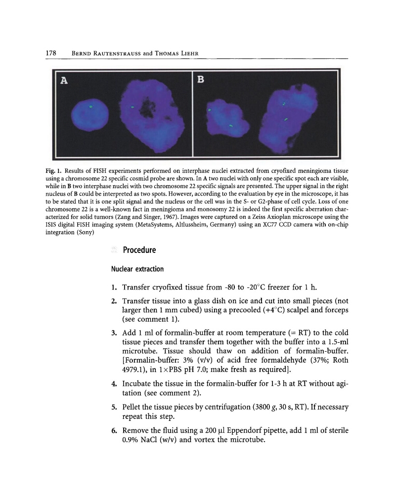
Fig. 1. Results of FISH experiments performed on interphase nuclei extracted from cryofIxed meningioma tissue
using a chromosome 22 specifIc cosmid probe are shown. In A two nuclei with only one specifIc spot each are visible,
while in B two interphase nuclei with two chromosome 22 specific signals are presented. The upper signal in the right
nucleus of B could be interpreted as two spots. However, according to the evaluation by eye in the microscope, it has
to be stated that it is one split signal and the nucleus or the cell was in the S- or G2-phase of cell cycle. Loss of one
chromosome 22 is a well-known fact in meningioma and monosomy 22 is indeed the first specific aberration characterized for solid tumors (Zang and Singer, 1967). Images were captured on a Zeiss Axioplan microscope using the
ISIS digital FISH imaging system (MetaSystems, Altlussheim, Germany) using an XC77 CCD camera with on-chip
integration (Sony)
Procedure
Nuclear extraction
1. Transfer cryofIxed tissue from -80 to -20°C freezer for 1 h.
2. Transfer tissue into a glass dish on ice and cut into small pieces (not
larger then 1 mm cubed) using a precooled (+4°C) scalpel and forceps
(see comment 1).
3. Add 1 ml of formalin-buffer at room temperature (= RT) to the cold
tissue pieces and transfer them together with the buffer into a 1.5-ml
microtube. Tissue should thaw on addition of formalin-buffer.
[Formalin-buffer: 3% (vlv) of acid free formaldehyde (37%; Roth
4979.1), in 1xPBS pH 7.0; make fresh as required].
4. Incubate the tissue in the formalin-buffer for 1-3 h at RT without agitation (see comment 2).
5. Pellet the tissue pieces by centrifugation (3800 g, 30 s, RT). If necessary
repeat this step.
6. Remove the fluid using a 200 fJI Eppendorf pipette, add 1 ml of sterile
0.9% NaCI (w/v) and vortex the microtube.
BERND RAUTENSTRAUSS and THOMAS LIEHR
Fig. 1. Results of FISH experiments performed on interphase nuclei extracted from cryofIxed meningioma tissue
using a chromosome 22 specifIc cosmid probe are shown. In A two nuclei with only one specifIc spot each are visible,
while in B two interphase nuclei with two chromosome 22 specific signals are presented. The upper signal in the right
nucleus of B could be interpreted as two spots. However, according to the evaluation by eye in the microscope, it has
to be stated that it is one split signal and the nucleus or the cell was in the S- or G2-phase of cell cycle. Loss of one
chromosome 22 is a well-known fact in meningioma and monosomy 22 is indeed the first specific aberration characterized for solid tumors (Zang and Singer, 1967). Images were captured on a Zeiss Axioplan microscope using the
ISIS digital FISH imaging system (MetaSystems, Altlussheim, Germany) using an XC77 CCD camera with on-chip
integration (Sony)
Procedure
Nuclear extraction
1. Transfer cryofIxed tissue from -80 to -20°C freezer for 1 h.
2. Transfer tissue into a glass dish on ice and cut into small pieces (not
larger then 1 mm cubed) using a precooled (+4°C) scalpel and forceps
(see comment 1).
3. Add 1 ml of formalin-buffer at room temperature (= RT) to the cold
tissue pieces and transfer them together with the buffer into a 1.5-ml
microtube. Tissue should thaw on addition of formalin-buffer.
[Formalin-buffer: 3% (vlv) of acid free formaldehyde (37%; Roth
4979.1), in 1xPBS pH 7.0; make fresh as required].
4. Incubate the tissue in the formalin-buffer for 1-3 h at RT without agitation (see comment 2).
5. Pellet the tissue pieces by centrifugation (3800 g, 30 s, RT). If necessary
repeat this step.
6. Remove the fluid using a 200 fJI Eppendorf pipette, add 1 ml of sterile
0.9% NaCI (w/v) and vortex the microtube.