

12 Nucleus Extraction from Formalin Fixed/paraffin Embedded Tissue
163
mounted (Liehr et al. 1995a, 1997) and from mounted tissue sections
(Liehr et al. 1999) is described.
Outline
1. Formalin-fIxed/paraffin-embedded tissue is cut into sections, which
are deparaffInized, rehydrated and treated with proteinase K to digest
the cytoplasm and set the nuclei free. This can be performed with
mounted and unmounted sections. Interphase nuclei are separated
from debris by passing through a nylon mesh. Nuclei are washed in
PBS and placed on uncoated, clean slides and fIxed. Small pieces of
tissue (Sx2xO.s mm) or one mounted section 1 cm in diameter are
suffIcient to produce two to fIve slides with extracted nuclei, suitable
for interphase FISH analyses.
2. For a FISH approach, these slides are treated with RNase and pepsin
prior to denaturation.
3. Denaturation of the slides is performed for about 12 min at 75°C. Biotinylated and digoxigenated probes are denatured for 5 min and hybridized on the slides for 3 nights at 37°C in a humid chamber. After a
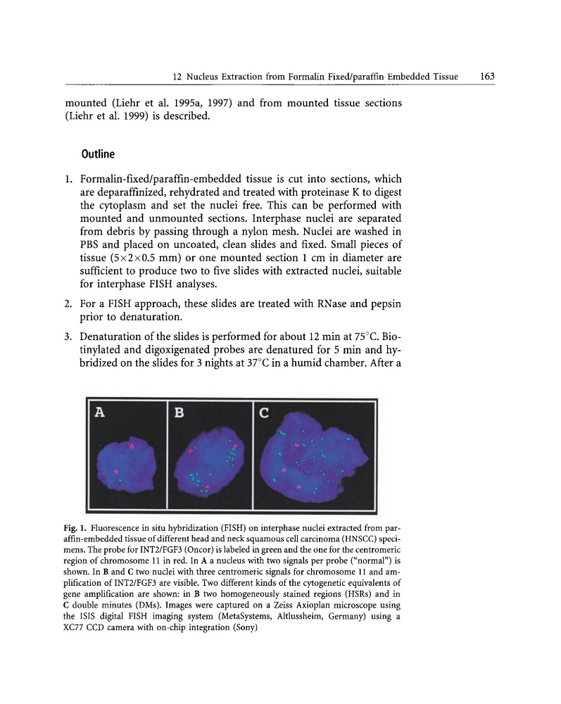
Fig. I. Fluorescence in situ hybridization (FISH) on interphase nuclei extracted from paraffin-embedded tissue of different head and neck squamous cell carcinoma (HNSCC) specimens. The probe for INT2/FGF3 (Oncor) is labeled in green and the one for the centromeric
region of chromosome 11 in red. In A a nucleus with two signals per probe ("normal") is
shown. In Band C two nuclei with three centromeric signals for chromosome 11 and amplification of INT2/FGF3 are visible. Two different kinds of the cytogenetic equivalents of
gene amplification are shown: in B two homogeneously stained regions (HSRs) and in
C double minutes (DMs). Images were captured on a Zeiss Axioplan microscope using
the ISIS digital FISH imaging system (MetaSystems, Altlussheim, Germany) using a
XC77 CCD camera with on-chip integration (Sony)
163
mounted (Liehr et al. 1995a, 1997) and from mounted tissue sections
(Liehr et al. 1999) is described.
Outline
1. Formalin-fIxed/paraffin-embedded tissue is cut into sections, which
are deparaffInized, rehydrated and treated with proteinase K to digest
the cytoplasm and set the nuclei free. This can be performed with
mounted and unmounted sections. Interphase nuclei are separated
from debris by passing through a nylon mesh. Nuclei are washed in
PBS and placed on uncoated, clean slides and fIxed. Small pieces of
tissue (Sx2xO.s mm) or one mounted section 1 cm in diameter are
suffIcient to produce two to fIve slides with extracted nuclei, suitable
for interphase FISH analyses.
2. For a FISH approach, these slides are treated with RNase and pepsin
prior to denaturation.
3. Denaturation of the slides is performed for about 12 min at 75°C. Biotinylated and digoxigenated probes are denatured for 5 min and hybridized on the slides for 3 nights at 37°C in a humid chamber. After a
Fig. I. Fluorescence in situ hybridization (FISH) on interphase nuclei extracted from paraffin-embedded tissue of different head and neck squamous cell carcinoma (HNSCC) specimens. The probe for INT2/FGF3 (Oncor) is labeled in green and the one for the centromeric
region of chromosome 11 in red. In A a nucleus with two signals per probe ("normal") is
shown. In Band C two nuclei with three centromeric signals for chromosome 11 and amplification of INT2/FGF3 are visible. Two different kinds of the cytogenetic equivalents of
gene amplification are shown: in B two homogeneously stained regions (HSRs) and in
C double minutes (DMs). Images were captured on a Zeiss Axioplan microscope using
the ISIS digital FISH imaging system (MetaSystems, Altlussheim, Germany) using a
XC77 CCD camera with on-chip integration (Sony)