

6 Epithelial Cells from Buccal Smears and Urine
107
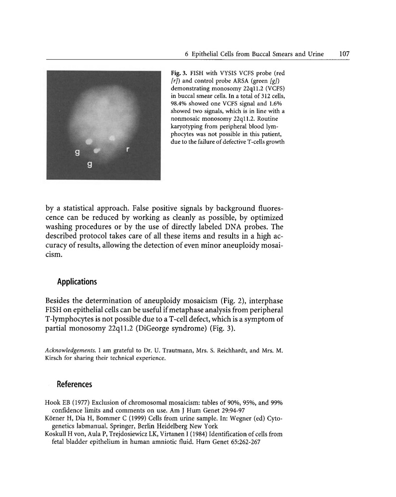
Fig. 3. FISH with VYSIS VCFS probe (red
[rJ) and control probe ARSA (green [gJ)
demonstrating monosomy 22ql1.2 (VCFS)
in buccal smear cells. In a total of 3I2 cells,
98.4% showed one VCFS signal and 1.6%
showed two signals, which is in line with a
nonmosaic monosomy 22q11.2. Routine
karyotyping from peripheral blood lymphocytes was not possible in this patient,
due to the failure of defective T-cells growth
by a statistical approach. False positive signals by background fluorescence can be reduced by working as cleanly as possible, by optimized
washing procedures or by the use of directly labeled DNA probes. The
described protocol takes care of all these items and results in a high accuracy of results, allowing the detection of even minor aneuploidy mosaicism.
Applications
Besides the determination of aneuploidy mosaicism (Fig. 2), interphase
FISH on epithelial cells can be useful ifmetaphase analysis from peripheral
T-lymphocytes is not possible due to aT-cell defect, which is a symptom of
partial monosomy 22ql1.2 (DiGeorge syndrome) (Fig. 3).
Acknowledgements. I am grateful to Dr. U. Trautmann, Mrs. S. Reichhardt, and Mrs. M.
Kirsch for sharing their technical experience.
References
Hook EB (1977) Exclusion of chromosomal mosaicism: tables of 90%, 95%. and 99%
confidence limits and comments on use. Am J Hum Genet 29:94-97
Korner H, Dia H, Bommer C (1999) Cells from urine sample. In: Wegner (ed) Cytogenetics labmanual. Springer, Berlin Heidelberg New York
Koskull H von, Aula P, Trejdosiewicz LK, Virtanen I (1984) Identification of cells from
fetal bladder epithelium in human amniotic fluid. Hum Genet 65:262-267
107
Fig. 3. FISH with VYSIS VCFS probe (red
[rJ) and control probe ARSA (green [gJ)
demonstrating monosomy 22ql1.2 (VCFS)
in buccal smear cells. In a total of 3I2 cells,
98.4% showed one VCFS signal and 1.6%
showed two signals, which is in line with a
nonmosaic monosomy 22q11.2. Routine
karyotyping from peripheral blood lymphocytes was not possible in this patient,
due to the failure of defective T-cells growth
by a statistical approach. False positive signals by background fluorescence can be reduced by working as cleanly as possible, by optimized
washing procedures or by the use of directly labeled DNA probes. The
described protocol takes care of all these items and results in a high accuracy of results, allowing the detection of even minor aneuploidy mosaicism.
Applications
Besides the determination of aneuploidy mosaicism (Fig. 2), interphase
FISH on epithelial cells can be useful ifmetaphase analysis from peripheral
T-lymphocytes is not possible due to aT-cell defect, which is a symptom of
partial monosomy 22ql1.2 (DiGeorge syndrome) (Fig. 3).
Acknowledgements. I am grateful to Dr. U. Trautmann, Mrs. S. Reichhardt, and Mrs. M.
Kirsch for sharing their technical experience.
References
Hook EB (1977) Exclusion of chromosomal mosaicism: tables of 90%, 95%. and 99%
confidence limits and comments on use. Am J Hum Genet 29:94-97
Korner H, Dia H, Bommer C (1999) Cells from urine sample. In: Wegner (ed) Cytogenetics labmanual. Springer, Berlin Heidelberg New York
Koskull H von, Aula P, Trejdosiewicz LK, Virtanen I (1984) Identification of cells from
fetal bladder epithelium in human amniotic fluid. Hum Genet 65:262-267