

5 FISH on Blood or Bone Marrow Smears
95
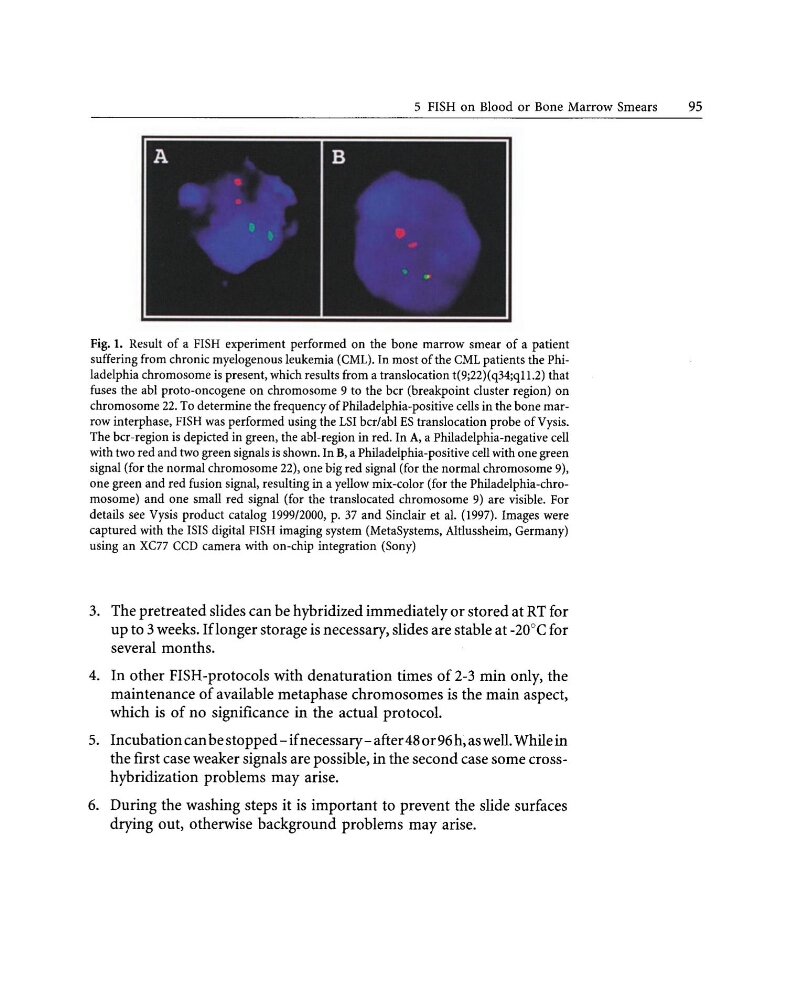
Fig. 1. Result of a FISH experiment performed on the bone marrow smear of a patient
suffering from chronic myelogenous leukemia (CML). In most of the CML patients the Philadelphia chromosome is present, which results from a translocation t(9;22)(q34;ql 1.2) that
fuses the abl proto-oncogene on chromosome 9 to the bcr (breakpoint cluster region) on
chromosome 22. To determine the frequency of Philadelphia-positive cells in the bone marrow interphase, FISH was performed using the LSI bcr/abl ES translocation probe ofVysis.
The bcr-region is depicted in green, the abl-region in red. In A, a Philadelphia-negative cell
with two red and two green signals is shown. In B, a Philadelphia-positive cell with one green
signal (for the normal chromosome 22), one big red signal (for the normal chromosome 9),
one green and red fusion signal, resulting in a yellow mix-color (for the Philadelphia-chromosome) and one small red signal (for the translocated chromosome 9) are visible. For
details see Vysis product catalog 1999/2000, p. 37 and Sinclair et al. (1997). Images were
captured with the ISIS digital FISH imaging system (MetaSystems, Altlussheim, Germany)
using an xcn CCD camera with on-chip integration (Sony)
3. The pretreated slides can be hybridized immediately or stored at RT for
up to 3 weeks. Iflonger storage is necessary, slides are stable at -20°C for
several months.
4. In other FISH-protocols with denaturation times of 2-3 min only, the
maintenance of available metaphase chromosomes is the main aspect,
which is of no significance in the actual protocol.
5. Incubation can bestopped-ifnecessary-after480r96h,aswell. While in
the first case weaker signals are possible, in the second case some crosshybridization problems may arise.
6. During the washing steps it is important to prevent the slide surfaces
drying out, otherwise background problems may arise.
95
Fig. 1. Result of a FISH experiment performed on the bone marrow smear of a patient
suffering from chronic myelogenous leukemia (CML). In most of the CML patients the Philadelphia chromosome is present, which results from a translocation t(9;22)(q34;ql 1.2) that
fuses the abl proto-oncogene on chromosome 9 to the bcr (breakpoint cluster region) on
chromosome 22. To determine the frequency of Philadelphia-positive cells in the bone marrow interphase, FISH was performed using the LSI bcr/abl ES translocation probe ofVysis.
The bcr-region is depicted in green, the abl-region in red. In A, a Philadelphia-negative cell
with two red and two green signals is shown. In B, a Philadelphia-positive cell with one green
signal (for the normal chromosome 22), one big red signal (for the normal chromosome 9),
one green and red fusion signal, resulting in a yellow mix-color (for the Philadelphia-chromosome) and one small red signal (for the translocated chromosome 9) are visible. For
details see Vysis product catalog 1999/2000, p. 37 and Sinclair et al. (1997). Images were
captured with the ISIS digital FISH imaging system (MetaSystems, Altlussheim, Germany)
using an xcn CCD camera with on-chip integration (Sony)
3. The pretreated slides can be hybridized immediately or stored at RT for
up to 3 weeks. Iflonger storage is necessary, slides are stable at -20°C for
several months.
4. In other FISH-protocols with denaturation times of 2-3 min only, the
maintenance of available metaphase chromosomes is the main aspect,
which is of no significance in the actual protocol.
5. Incubation can bestopped-ifnecessary-after480r96h,aswell. While in
the first case weaker signals are possible, in the second case some crosshybridization problems may arise.
6. During the washing steps it is important to prevent the slide surfaces
drying out, otherwise background problems may arise.