

250
G. Tagliafierro et al.
•
' .
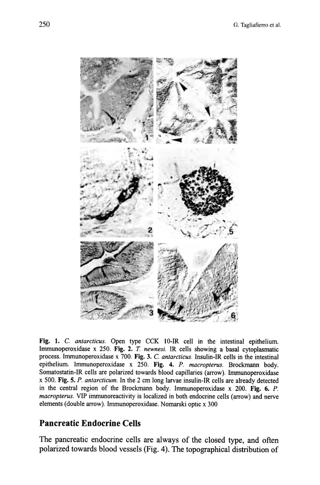
Fig. 1. C. antarcticus. Open type CCK lO-IR cell in the intestinal epithelium.
Immunoperoxidase x 250. Fig. 2. T newnesi. IR cells showing a basal cytoplasmatic
process. Immunoperoxidase x 700. Fig. 3. C. antarcticus. Insulin-IR cells in the intestinal
epithelium. Immunoperoxidase x 250. Fig. 4. P. macropterus. Brockmann body.
Somatostatin-IR cells are polarized towards blood capillaries (arrow). Immunoperoxidase
x 500. Fig. 5. P. antarcticum. In the 2 cm long larvae insulin-IR cells are already detected
in the central region of the Brockmann body. Immunoperoxidase x 200. Fig. 6. P.
macropterus. VIP immunoreactivity is localized in both endocrine cells (arrow) and nerve
elements (double arrow). Immunoperoxidase. Nomarski optic x 300
Pancreatic Endocrine Cells
The pancreatic endocrine cells are always of the closed type, and often
polarized towards blood vessels (Fig. 4). The topographical distribution of
G. Tagliafierro et al.
•
' .
Fig. 1. C. antarcticus. Open type CCK lO-IR cell in the intestinal epithelium.
Immunoperoxidase x 250. Fig. 2. T newnesi. IR cells showing a basal cytoplasmatic
process. Immunoperoxidase x 700. Fig. 3. C. antarcticus. Insulin-IR cells in the intestinal
epithelium. Immunoperoxidase x 250. Fig. 4. P. macropterus. Brockmann body.
Somatostatin-IR cells are polarized towards blood capillaries (arrow). Immunoperoxidase
x 500. Fig. 5. P. antarcticum. In the 2 cm long larvae insulin-IR cells are already detected
in the central region of the Brockmann body. Immunoperoxidase x 200. Fig. 6. P.
macropterus. VIP immunoreactivity is localized in both endocrine cells (arrow) and nerve
elements (double arrow). Immunoperoxidase. Nomarski optic x 300
Pancreatic Endocrine Cells
The pancreatic endocrine cells are always of the closed type, and often
polarized towards blood vessels (Fig. 4). The topographical distribution of