

Streptocephalus texan us Packard, 1871
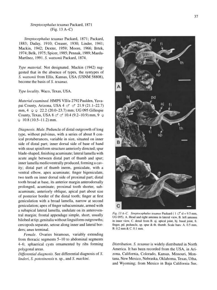
(Fig. 13 A-C)
Streptocephalus texanus Packard, 1871; Packard,
1883; Daday, 1910; Creaser, 1930; Linder, 1941;
Mackin, 1942; Dexter, 1959; Moore, 1966; Brtek,
1974; Belk, 1975; Spicer, 1985; Pennak, 1989; MaedaMartinez, 1991. S. watsonii Packard, 1874.
Type material. Not designated. Mackin (1942) suggested that in the absence of types, the syntypes of
S. watsonii from Ellis, Kansas, USA (USNM 58808),
become the basis of S. texanus.
Type locality. Waco, Texas, USA.
Material examined. HMPS VII/a-2792 Paulden, Yavapai County, Arizona, USA 4 d d 21.9 (21.1-22.7)
mm,4 Q Q 22.2 (20.0-23.7) mm; UG 095 Gillespie
County, Texas, USA 8 d d lOA (9.2-10.9) mm, 9 Q
Q 10.8 (10.5-11.2) mm.
Diagnosis. Male. Peduncle of distal outgrowth of long
type, without pulvinus, with a series of about 8 conical protuberances, variable in size, situated on inner
side of distal part; inner dorsal side of base of hand
with stout spiniform structure anteriorly directed; spur
blade-shaped, finishing acuminate; lateral lamella with
acute angle between distal part of thumb and spur;
inner lamella medioventrally produced, forming a cavity; distal part of thumb inerm, geniculate, with a
ventral elbow, apex acuminate; finger bigeniculate,
two teeth on inner dorsal side of proximal part; distal
tooth broad at base, its anterior margin anterodorsally
prolonged, acuminate; proximal tooth shorter, subacuminate, anteriorly oblique, apical part about size
of posterior border of the distal tooth; finger at first
geniculation with a broad lamella, narrow at second
geniculation; apex of finger subacuminate, armed with
a subapical lateral lamella, undulate on its anteroventral margin ; frontal appendage simple, short, usually
bilobed at tip; genitalia without linguiform outgrowths;
cercopods separate, setose along inner and lateral borders; anus terminal.
Female . Ovaries biramous, variably extending
from thoracic segments 5-10 to abdominal segments
4-6; sphaerical cysts ornamented by ribs forming
polygonal areas.
Differential diagnosis. See differential diagnosis of S.
linderi , S. potosinensis n. sp., and S. mackini.
37
Fig. 13 A-C. Streptocephalus texan us Packard ( I d sl = 9.5 mm,
UG 095). A. Head and right antenna in lateral view, B. left antenna
in inner view, C. detail from B. aj. apical joint, bj. basal joint, fi.
finger, pd. peduncle, sp. spur & tho thumb. Scale bars: A. 0.5 mm,
B. 0.2 mm & C. 0.1 mm.
Distribution. S. texanus is widely distributed in North
America. It has been recorded from the USA, in Arizona, California, Colorado, Kansas, Missouri, Montana, New Mexico, Nebraska, Oklahoma, Texas, Utha,
and Wyoming; from Mexico in Baja California Sur,
(Fig. 13 A-C)
Streptocephalus texanus Packard, 1871; Packard,
1883; Daday, 1910; Creaser, 1930; Linder, 1941;
Mackin, 1942; Dexter, 1959; Moore, 1966; Brtek,
1974; Belk, 1975; Spicer, 1985; Pennak, 1989; MaedaMartinez, 1991. S. watsonii Packard, 1874.
Type material. Not designated. Mackin (1942) suggested that in the absence of types, the syntypes of
S. watsonii from Ellis, Kansas, USA (USNM 58808),
become the basis of S. texanus.
Type locality. Waco, Texas, USA.
Material examined. HMPS VII/a-2792 Paulden, Yavapai County, Arizona, USA 4 d d 21.9 (21.1-22.7)
mm,4 Q Q 22.2 (20.0-23.7) mm; UG 095 Gillespie
County, Texas, USA 8 d d lOA (9.2-10.9) mm, 9 Q
Q 10.8 (10.5-11.2) mm.
Diagnosis. Male. Peduncle of distal outgrowth of long
type, without pulvinus, with a series of about 8 conical protuberances, variable in size, situated on inner
side of distal part; inner dorsal side of base of hand
with stout spiniform structure anteriorly directed; spur
blade-shaped, finishing acuminate; lateral lamella with
acute angle between distal part of thumb and spur;
inner lamella medioventrally produced, forming a cavity; distal part of thumb inerm, geniculate, with a
ventral elbow, apex acuminate; finger bigeniculate,
two teeth on inner dorsal side of proximal part; distal
tooth broad at base, its anterior margin anterodorsally
prolonged, acuminate; proximal tooth shorter, subacuminate, anteriorly oblique, apical part about size
of posterior border of the distal tooth; finger at first
geniculation with a broad lamella, narrow at second
geniculation; apex of finger subacuminate, armed with
a subapical lateral lamella, undulate on its anteroventral margin ; frontal appendage simple, short, usually
bilobed at tip; genitalia without linguiform outgrowths;
cercopods separate, setose along inner and lateral borders; anus terminal.
Female . Ovaries biramous, variably extending
from thoracic segments 5-10 to abdominal segments
4-6; sphaerical cysts ornamented by ribs forming
polygonal areas.
Differential diagnosis. See differential diagnosis of S.
linderi , S. potosinensis n. sp., and S. mackini.
37
Fig. 13 A-C. Streptocephalus texan us Packard ( I d sl = 9.5 mm,
UG 095). A. Head and right antenna in lateral view, B. left antenna
in inner view, C. detail from B. aj. apical joint, bj. basal joint, fi.
finger, pd. peduncle, sp. spur & tho thumb. Scale bars: A. 0.5 mm,
B. 0.2 mm & C. 0.1 mm.
Distribution. S. texanus is widely distributed in North
America. It has been recorded from the USA, in Arizona, California, Colorado, Kansas, Missouri, Montana, New Mexico, Nebraska, Oklahoma, Texas, Utha,
and Wyoming; from Mexico in Baja California Sur,