

26
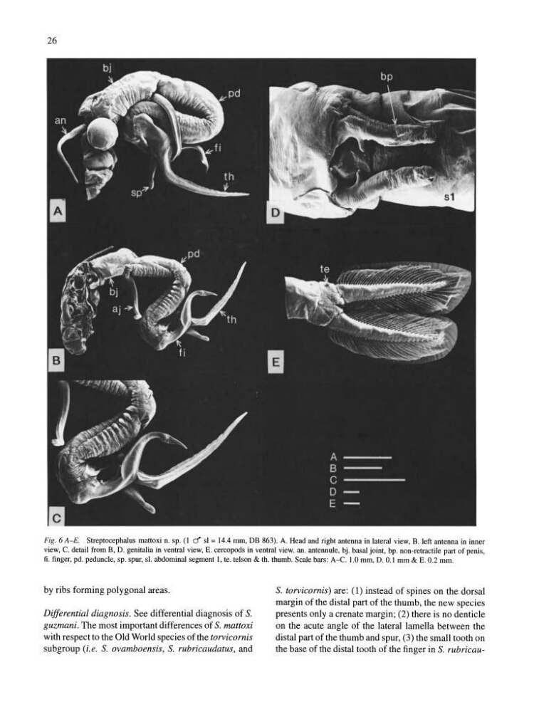
Fig. 6 A-E. Streptocephalus mattoxi n. sp. (1 d' sl = 14.4 mm, DB 863). A. Head and right antenna in lateral view, B. left antenna in inner
view, C. detail from B, D. genitalia in ventral view, E. cercopods in ventral view. an. antennule, bj. basal joint, bp. non-retractile part of penis,
fi. finger, pd. peduncle, sp. spur, sl. abdominal segment 1, teo telson & tho thumb. Scale bars: A-C. 1.0 mm, D. 0.1 mm & E. 0.2 mm.
by ribs forming polygonal areas.
Differential diagnosis. See differential diagnosis of S.
guzmani. The most important differences of S. mattoxi
with respect to the Old World species of the torvicornis
subgroup (i.e. S. ovamboensis, S. rubricaudatus, and
S. torvicornis) are: (1) instead of spines on the dorsal
margin of the distal part of the thumb, the new species
presents only a crenate margin; (2) there is no denticle
on the acute angle of the lateral lamella between the
distal part ofthe thumb and spur, (3) the small tooth on
the base of the distal tooth of the finger in S. rubricau-
Fig. 6 A-E. Streptocephalus mattoxi n. sp. (1 d' sl = 14.4 mm, DB 863). A. Head and right antenna in lateral view, B. left antenna in inner
view, C. detail from B, D. genitalia in ventral view, E. cercopods in ventral view. an. antennule, bj. basal joint, bp. non-retractile part of penis,
fi. finger, pd. peduncle, sp. spur, sl. abdominal segment 1, teo telson & tho thumb. Scale bars: A-C. 1.0 mm, D. 0.1 mm & E. 0.2 mm.
by ribs forming polygonal areas.
Differential diagnosis. See differential diagnosis of S.
guzmani. The most important differences of S. mattoxi
with respect to the Old World species of the torvicornis
subgroup (i.e. S. ovamboensis, S. rubricaudatus, and
S. torvicornis) are: (1) instead of spines on the dorsal
margin of the distal part of the thumb, the new species
presents only a crenate margin; (2) there is no denticle
on the acute angle of the lateral lamella between the
distal part ofthe thumb and spur, (3) the small tooth on
the base of the distal tooth of the finger in S. rubricau-