

Hydrobiologia 298: 183-201, 1995.
D. Belk, H. 1. Dumont & G. Maier (eds), Studies on Large Branchiopod Biology and Aquaculture II.
© 1995 Kluwer Academic Publishers.
183
An analysis of the setation pattern of the limbs in Anostraca (Crustacea);
using the Algerian species as an example
Lynda Beladjal 1 , Johan Mertens 2 ,* & Henri J. Dumont 2
1 Lab. Ecologie Animale, Univ. S. T.H.B., BP 32 EI Alia - Bab Ezzouar, Alger, Algeria
2 Laboratory of Animal Ecology, University of Ghent, K. L. Ledeganckstraat 35, B-9000 Ghent, Belgium
(* author for correspondence)
Key words: Anostraca, limb structure, Algeria
Abstract
The implantation, number, and structure of the setae on the endopodites and exopodites of the limbs, and the
setulation of the setae of endite I of all Anostracan species found in Algeria are analysed by light and scanning
electron microscopy and graphically represented. The data disclose significant variation among the genera A rtemia ,
Branchinella, Branchipus, Chirocephalus, Streptocephalus and Tanymastigites, and between the related congeners
Streptocephalus torvicornis and S. rubricaudatus.
Introduction
Recently, we described the limb structure of some
Streptocephalus, based on scanning electron micrographs (Mertens et al., 1991), demonstrating morphological differences among three species in the fine
structure of the setae on the endites. We explored the
possibility that such species-specific differences of the
filtration apparatus might contribute to selectivity in
feeding. Here, we show that quite a few unique quantitative and qualitative characters of the endite I and of
the endo- and exopodites have been ignored since Linder (1941) proclaimed that no relevant differences in
limb structure occur in the Anostraca. We also demonstrate that these structures are usable as a taxonomic
tool at the genus - and perhaps at the species level.
Materials and methods
All material investigated here was obtained from Anostraca collected in Algeria in their natural habitats, by
the authors (Table 1). Limbs of full-grown males and
females of each species were dissected and mounted
for a study by light microscopy, or critical-point dried,
and gold coated for SEM-work.
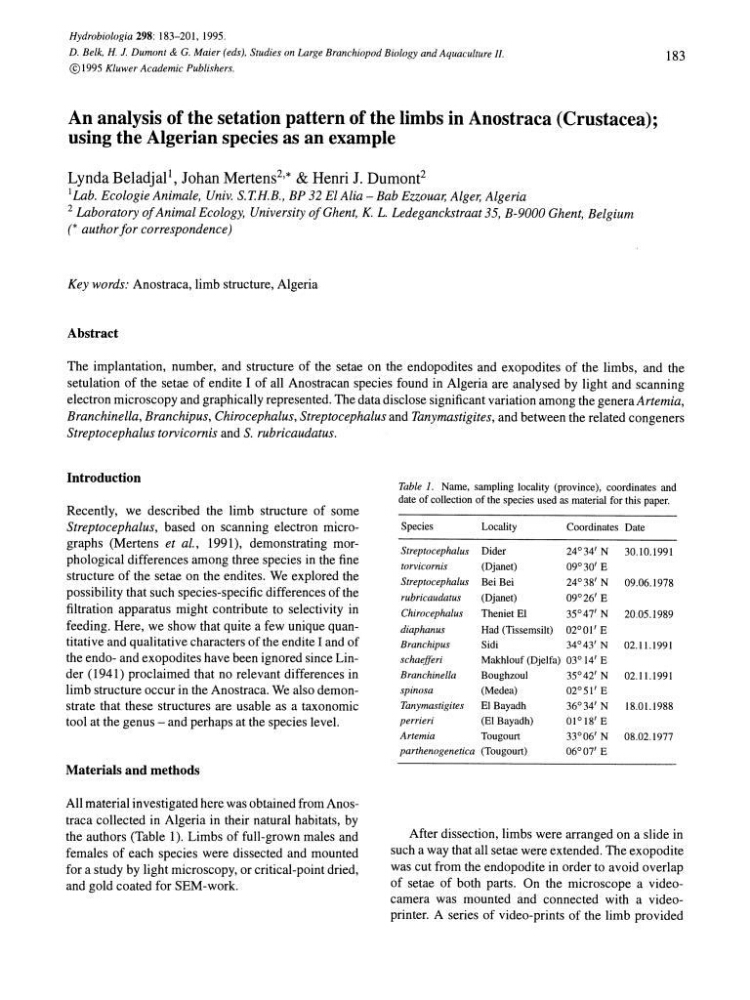
Table 1. Name, sampling locality (province), coordinates and
date of collection of the species used as material for this paper.
Species
Locality
Coordinates Date
Streptocephalus Dider
24°34' N
30.10.1991
torvicornis
(Djanet)
09°30' E
Streptocephalus Bei Bei
24°38' N
09.06.1978
rubricaudatus
(Djanet)
09°26' E
Chirocephalus
Theniet El
35°47' N
20.05.1989
diaphanus
Had (Tissemsilt) 02°01' E
Branchipus
Sidi
34°43' N 02.11.1991
schaefferi
Makh10uf (Djelfa) 03° 14' E
Branchinella
Boughzoul
35°42' N 02.1l.l991
spinosa
(Medea)
02°51' E
Tanymastigites
EI Bayadh
36°34' N
18.01.1988
perrieri
(El Bayadh)
01°18'E
Artemia
Tougourt
33°06' N 08.02.1977
parthenogenetica (Tougourt)
06°07' E
After dissection, limbs were arranged on a slide in
such a way that all setae were extended. The exopodite
was cut from the endopodite in order to avoid overlap
of setae of both parts. On the microscope a videocamera was mounted and connected with a videoprinter. A series of video-prints of the limb provided
D. Belk, H. 1. Dumont & G. Maier (eds), Studies on Large Branchiopod Biology and Aquaculture II.
© 1995 Kluwer Academic Publishers.
183
An analysis of the setation pattern of the limbs in Anostraca (Crustacea);
using the Algerian species as an example
Lynda Beladjal 1 , Johan Mertens 2 ,* & Henri J. Dumont 2
1 Lab. Ecologie Animale, Univ. S. T.H.B., BP 32 EI Alia - Bab Ezzouar, Alger, Algeria
2 Laboratory of Animal Ecology, University of Ghent, K. L. Ledeganckstraat 35, B-9000 Ghent, Belgium
(* author for correspondence)
Key words: Anostraca, limb structure, Algeria
Abstract
The implantation, number, and structure of the setae on the endopodites and exopodites of the limbs, and the
setulation of the setae of endite I of all Anostracan species found in Algeria are analysed by light and scanning
electron microscopy and graphically represented. The data disclose significant variation among the genera A rtemia ,
Branchinella, Branchipus, Chirocephalus, Streptocephalus and Tanymastigites, and between the related congeners
Streptocephalus torvicornis and S. rubricaudatus.
Introduction
Recently, we described the limb structure of some
Streptocephalus, based on scanning electron micrographs (Mertens et al., 1991), demonstrating morphological differences among three species in the fine
structure of the setae on the endites. We explored the
possibility that such species-specific differences of the
filtration apparatus might contribute to selectivity in
feeding. Here, we show that quite a few unique quantitative and qualitative characters of the endite I and of
the endo- and exopodites have been ignored since Linder (1941) proclaimed that no relevant differences in
limb structure occur in the Anostraca. We also demonstrate that these structures are usable as a taxonomic
tool at the genus - and perhaps at the species level.
Materials and methods
All material investigated here was obtained from Anostraca collected in Algeria in their natural habitats, by
the authors (Table 1). Limbs of full-grown males and
females of each species were dissected and mounted
for a study by light microscopy, or critical-point dried,
and gold coated for SEM-work.
Table 1. Name, sampling locality (province), coordinates and
date of collection of the species used as material for this paper.
Species
Locality
Coordinates Date
Streptocephalus Dider
24°34' N
30.10.1991
torvicornis
(Djanet)
09°30' E
Streptocephalus Bei Bei
24°38' N
09.06.1978
rubricaudatus
(Djanet)
09°26' E
Chirocephalus
Theniet El
35°47' N
20.05.1989
diaphanus
Had (Tissemsilt) 02°01' E
Branchipus
Sidi
34°43' N 02.11.1991
schaefferi
Makh10uf (Djelfa) 03° 14' E
Branchinella
Boughzoul
35°42' N 02.1l.l991
spinosa
(Medea)
02°51' E
Tanymastigites
EI Bayadh
36°34' N
18.01.1988
perrieri
(El Bayadh)
01°18'E
Artemia
Tougourt
33°06' N 08.02.1977
parthenogenetica (Tougourt)
06°07' E
After dissection, limbs were arranged on a slide in
such a way that all setae were extended. The exopodite
was cut from the endopodite in order to avoid overlap
of setae of both parts. On the microscope a videocamera was mounted and connected with a videoprinter. A series of video-prints of the limb provided