

17 Effect of Dilution Rate on Azadinium spinosum and Azaspiracid (AZA). . .
199
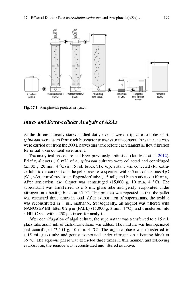
Fig. 17.1 Azaspiracids production system
Intra- and Extra-cellular Analysis of AZAs
At the different steady states studied daily over a week, triplicate samples of A.
spinosum were taken from each bioreactor to assess toxin content, the same analyses
were carried out from the 300 L harvesting tank before each tangential flow filtration
for initial toxin content assessment.
The analytical procedure had been previously optimised (Jauffrais et al. 2012).
Briefly, aliquots (10 mL) of A. spinosum cultures were collected and centrifuged
(2,500 g, 20 min, 4
ı C) in 15 mL tubes. The supernatant was collected (for extracellular toxin content) and the pellet was re-suspended with 0.5 mL of acetone/H 2 O
(9/1, v/v), transferred to an Eppendorf tube (1.5 mL) and bath sonicated (10 min).
After sonication, the aliquot was centrifuged (15,000 g, 10 min, 4
ı C). The
supernatant was transferred to a 5 mL glass tube and gently evaporated under
nitrogen on a heating block at 35
ı C. This process was repeated so that the pellet
was extracted three times in total. After evaporation of supernatants, the residue
was reconstituted in 1 mL methanol. Subsequently, an aliquot was filtered with
NANOSEP MF filter 0.2 m (PALL) (15,000 g, 3 min, 4
ı C), and transferred into
a HPLC vial with a 250 L insert for analysis.
After centrifugation of algal culture, the supernatant was transferred to a 15 mL
glass tube and 5 mL of dichloromethane was added. The mixture was homogenized
and centrifuged (2,500 g, 10 min, 4
ı C). The organic phase was transferred to
a 15 mL glass tube and gently evaporated under nitrogen on a heating block at
35
ı C. The aqueous phase was extracted three times in this manner, and following
evaporation, the residue was reconstituted and filtered as above.
199
Fig. 17.1 Azaspiracids production system
Intra- and Extra-cellular Analysis of AZAs
At the different steady states studied daily over a week, triplicate samples of A.
spinosum were taken from each bioreactor to assess toxin content, the same analyses
were carried out from the 300 L harvesting tank before each tangential flow filtration
for initial toxin content assessment.
The analytical procedure had been previously optimised (Jauffrais et al. 2012).
Briefly, aliquots (10 mL) of A. spinosum cultures were collected and centrifuged
(2,500 g, 20 min, 4
ı C) in 15 mL tubes. The supernatant was collected (for extracellular toxin content) and the pellet was re-suspended with 0.5 mL of acetone/H 2 O
(9/1, v/v), transferred to an Eppendorf tube (1.5 mL) and bath sonicated (10 min).
After sonication, the aliquot was centrifuged (15,000 g, 10 min, 4
ı C). The
supernatant was transferred to a 5 mL glass tube and gently evaporated under
nitrogen on a heating block at 35
ı C. This process was repeated so that the pellet
was extracted three times in total. After evaporation of supernatants, the residue
was reconstituted in 1 mL methanol. Subsequently, an aliquot was filtered with
NANOSEP MF filter 0.2 m (PALL) (15,000 g, 3 min, 4
ı C), and transferred into
a HPLC vial with a 250 L insert for analysis.
After centrifugation of algal culture, the supernatant was transferred to a 15 mL
glass tube and 5 mL of dichloromethane was added. The mixture was homogenized
and centrifuged (2,500 g, 10 min, 4
ı C). The organic phase was transferred to
a 15 mL glass tube and gently evaporated under nitrogen on a heating block at
35
ı C. The aqueous phase was extracted three times in this manner, and following
evaporation, the residue was reconstituted and filtered as above.