

2 .2. THE CASE STUDIES
55
tentacl es
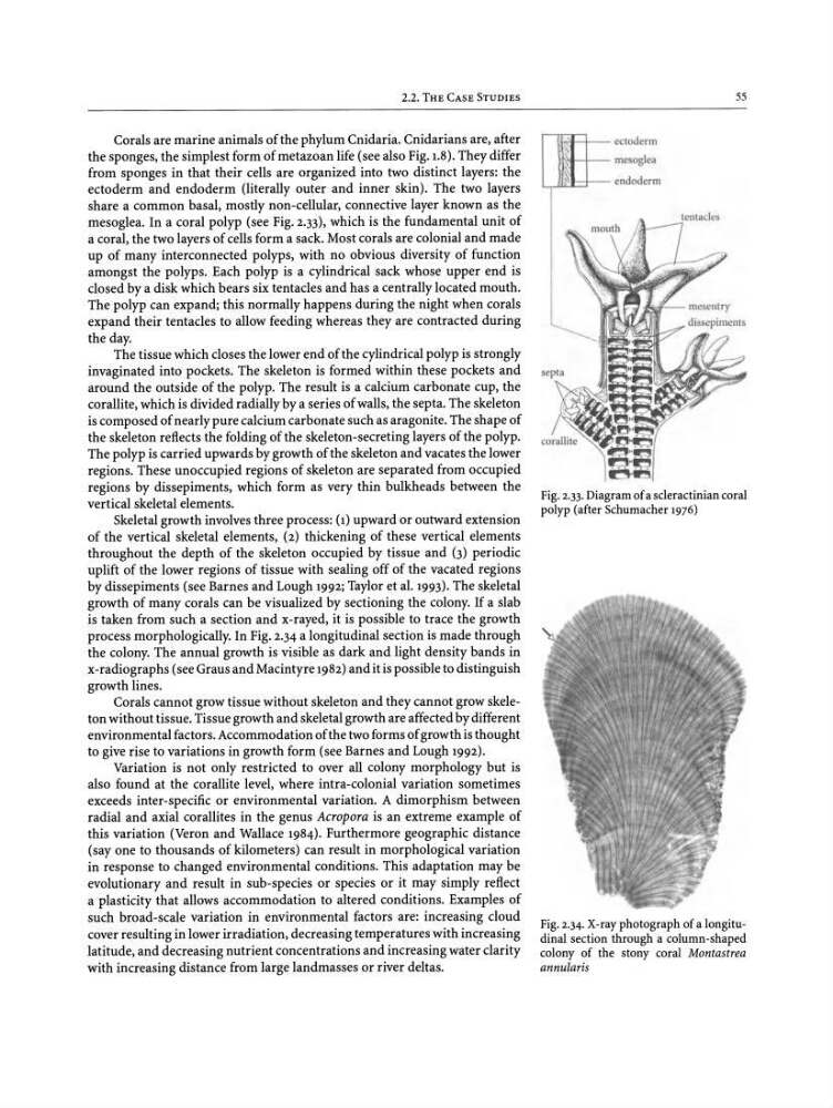
Fig. 2.33. Diagram ofa scleractinian coral
polyp(afterSchumacher 1976)
ectoderm
mesoglea
endoderm
'. ~
~ I- -
Fig. 2.34. X-ray photograph of a longitudinal section through a column-shaped
colony of the stony coral Montastrea
annularis
Corals are marine animals of the phylum Cnidaria. Cnidarians are, after
the sponges, the simplest form of metazoan life (see also Pig.j.s). They differ
from sponges in that their cells are organized into two distinct layers: the
ectoderm and endoderm (literally outer and inner skin). The two layers
share a common basal, mostly non -cellular, connective layer known as the
mesoglea. In a coral polyp (see Fig. 2.33), which is the fundamental unit of
a coral, the two layers of cells form a sack. Most corals are colonial and made
up of many interconnected polyps, with no obvious diversity of function
amongst the polyps . Each polyp is a cylindrical sack whose upper end is
closed by a disk which bears six tentacles and has a centrally located mouth.
The polyp can expand; this normally happens during the night when corals
expand their tentacles to allow feeding whereas they are contracted during
the day.
The tissue which closes the lower end of the cylindrical polyp is strongly
invaginated into pockets. The skeleton is formed within these pockets and
around the outside of the polyp. The result is a calcium carbonate cup, the
corallite, which is divided radially by a series of walls, the septa. The skeleton
is composed of nearly pure calcium carbonate such as aragonite. The shape of
the skeleton reflects the folding of the skeleton-secreting layers of the polyp.
The polyp is carried upwards by growth of the skeleton and vacates the lower
regions. These unoccupied regions of skeleton are separated from occupied
regions by dissepiments, which form as very thin bulkheads between the
vertical skeletal elements.
Skeletal growth involves three process: (1) upward or outward extension
of the vertical skeletal elements, (2) thickening of these vertical elements
throughout the depth of the skeleton occupied by tissue and (3) periodic
uplift of the lower regions of tissue with sealing off of the vacated regions
by dissepiments (see Barnes and Lough 1992; Taylor et al. 1993). The skeletal
growth of many corals can be visualized by sectioning the colony. If a slab
is taken from such a section and x-rayed, it is possible to trace the growth
process morphologically. In Fig. 2.34 a longitudinal section is made through
the colony. The annual growth is visible as dark and light density bands in
x-radiographs (see Graus and Macintyre 1982) and it is possible to distinguish
growth lines.
Corals cannot grow tissue without skeleton and they cannot grow skeleton without tissue. Tissue growth and skeletal growth are affected by different
environmental factors. Accommodation of the two forms of growth is thought
to give rise to variations in growth form (see Barnes and Lough 1992).
Variation is not only restricted to over all colony morphology but is
also found at the corallite level, where intra-colonial variation sometimes
exceeds inter-specific or environmental variation. A dimorphism between
radial and axial corallites in the genus Acropora is an extreme example of
this variation (Veron and Wallace 1984). Furthermore geographic distance
(say one to thousands of kilometers) can result in morphological variation
in response to changed environmental conditions. This adaptation may be
evolutionary and result in sub-species or species or it may simply reflect
a plasticity that allows accommodation to altered conditions. Examples of
such broad-scale variation in environmental factors are: increasing cloud
cover resulting in lower irradiation, decreasing temperatures with increasing
latitude, and decreasing nutrient concentrations and increasing water clarity
with increasing distance from large landmasses or river deltas.
55
tentacl es
Fig. 2.33. Diagram ofa scleractinian coral
polyp(afterSchumacher 1976)
ectoderm
mesoglea
endoderm
'. ~
~ I- -
Fig. 2.34. X-ray photograph of a longitudinal section through a column-shaped
colony of the stony coral Montastrea
annularis
Corals are marine animals of the phylum Cnidaria. Cnidarians are, after
the sponges, the simplest form of metazoan life (see also Pig.j.s). They differ
from sponges in that their cells are organized into two distinct layers: the
ectoderm and endoderm (literally outer and inner skin). The two layers
share a common basal, mostly non -cellular, connective layer known as the
mesoglea. In a coral polyp (see Fig. 2.33), which is the fundamental unit of
a coral, the two layers of cells form a sack. Most corals are colonial and made
up of many interconnected polyps, with no obvious diversity of function
amongst the polyps . Each polyp is a cylindrical sack whose upper end is
closed by a disk which bears six tentacles and has a centrally located mouth.
The polyp can expand; this normally happens during the night when corals
expand their tentacles to allow feeding whereas they are contracted during
the day.
The tissue which closes the lower end of the cylindrical polyp is strongly
invaginated into pockets. The skeleton is formed within these pockets and
around the outside of the polyp. The result is a calcium carbonate cup, the
corallite, which is divided radially by a series of walls, the septa. The skeleton
is composed of nearly pure calcium carbonate such as aragonite. The shape of
the skeleton reflects the folding of the skeleton-secreting layers of the polyp.
The polyp is carried upwards by growth of the skeleton and vacates the lower
regions. These unoccupied regions of skeleton are separated from occupied
regions by dissepiments, which form as very thin bulkheads between the
vertical skeletal elements.
Skeletal growth involves three process: (1) upward or outward extension
of the vertical skeletal elements, (2) thickening of these vertical elements
throughout the depth of the skeleton occupied by tissue and (3) periodic
uplift of the lower regions of tissue with sealing off of the vacated regions
by dissepiments (see Barnes and Lough 1992; Taylor et al. 1993). The skeletal
growth of many corals can be visualized by sectioning the colony. If a slab
is taken from such a section and x-rayed, it is possible to trace the growth
process morphologically. In Fig. 2.34 a longitudinal section is made through
the colony. The annual growth is visible as dark and light density bands in
x-radiographs (see Graus and Macintyre 1982) and it is possible to distinguish
growth lines.
Corals cannot grow tissue without skeleton and they cannot grow skeleton without tissue. Tissue growth and skeletal growth are affected by different
environmental factors. Accommodation of the two forms of growth is thought
to give rise to variations in growth form (see Barnes and Lough 1992).
Variation is not only restricted to over all colony morphology but is
also found at the corallite level, where intra-colonial variation sometimes
exceeds inter-specific or environmental variation. A dimorphism between
radial and axial corallites in the genus Acropora is an extreme example of
this variation (Veron and Wallace 1984). Furthermore geographic distance
(say one to thousands of kilometers) can result in morphological variation
in response to changed environmental conditions. This adaptation may be
evolutionary and result in sub-species or species or it may simply reflect
a plasticity that allows accommodation to altered conditions. Examples of
such broad-scale variation in environmental factors are: increasing cloud
cover resulting in lower irradiation, decreasing temperatures with increasing
latitude, and decreasing nutrient concentrations and increasing water clarity
with increasing distance from large landmasses or river deltas.