

40
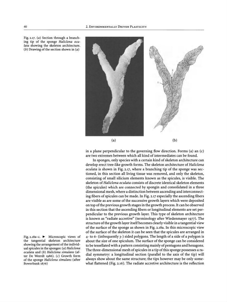
Fig. 2.17. (a) Section through a branching tip of the sponge Haliclona oculata showing the skeleton architecture.
(b) Drawing of the sectionshownin (a)
2. ENVIRONMENTALLY DRIVEN PLASTICITY
(a)
(b)
Fig. u8a-c." Microscopic views of
the tangential skeleton architecture
showing the arrangementof the individualspicules in the sponges: (a) Haliclona
oculata and (b) Haliclona simulans (after De Weerdt 1986). (c) Growth form
of the sponge Haliclona simulans (after
Bowerbank 1876)
in a plane perpendicular to the governing flow direction. Forms (a) an (c)
are two extremes between which all kind of intermediates can be found.
In sponges, only species with a certain kind of skeleton architecture can
develop erect tree-like growth forms. The skeleton architecture of Haliclona
oculata is shown in Fig. 2.17, where a branching tip of the sponge was sectioned, in this section all living tissue was removed, and only the skeleton ,
consisting of small silicium elements known as the spicules, is visible. The
skeleton of Haliclona oculata consists of discrete identical skeleton elements
(the spicules) which are connected by spongin and consolidated in a three
dimensional mesh, where a distinction between ascending and interconnecting fibers of spicules can be made. In Fig. 2.17 especially the ascending fibers
are visible as are some of the successive growth layers which were deposited
on top of the previous growth stages in the growth process . It can be observed
in this section that the ascending fibers or longitudinal elements are set perpendicular to the previous growth layer. This type of skeleton architecture
is known as "radiate accretive" (terminology after Wiedenmayer 1977). The
structure of the growth layer itselfbecomes clearly visible in a tangential view
of the surface of the sponge as shown in Fig. 2.18a. In this microscopic view
of the surface of the skeleton it can be seen that the spicules are arranged in
4- to 6- (infrequently 3-) sided polygons. The length of a side of a polygon is
about the size of one spiculum. The surface of the sponge can be considered
to be tessellated with a pattern consisting mainly of pentagons and hexagons.
The three-dimensional mesh of spicules in a tip of this sponge possesses a radial symmetry: a longitudinal section (parallel to the axis of the tip) will
always show about the same structure; the tips however may be only somewhat flattened (Fig. 2.16). The radiate accretive architecture is the reflection
Fig. 2.17. (a) Section through a branching tip of the sponge Haliclona oculata showing the skeleton architecture.
(b) Drawing of the sectionshownin (a)
2. ENVIRONMENTALLY DRIVEN PLASTICITY
(a)
(b)
Fig. u8a-c." Microscopic views of
the tangential skeleton architecture
showing the arrangementof the individualspicules in the sponges: (a) Haliclona
oculata and (b) Haliclona simulans (after De Weerdt 1986). (c) Growth form
of the sponge Haliclona simulans (after
Bowerbank 1876)
in a plane perpendicular to the governing flow direction. Forms (a) an (c)
are two extremes between which all kind of intermediates can be found.
In sponges, only species with a certain kind of skeleton architecture can
develop erect tree-like growth forms. The skeleton architecture of Haliclona
oculata is shown in Fig. 2.17, where a branching tip of the sponge was sectioned, in this section all living tissue was removed, and only the skeleton ,
consisting of small silicium elements known as the spicules, is visible. The
skeleton of Haliclona oculata consists of discrete identical skeleton elements
(the spicules) which are connected by spongin and consolidated in a three
dimensional mesh, where a distinction between ascending and interconnecting fibers of spicules can be made. In Fig. 2.17 especially the ascending fibers
are visible as are some of the successive growth layers which were deposited
on top of the previous growth stages in the growth process . It can be observed
in this section that the ascending fibers or longitudinal elements are set perpendicular to the previous growth layer. This type of skeleton architecture
is known as "radiate accretive" (terminology after Wiedenmayer 1977). The
structure of the growth layer itselfbecomes clearly visible in a tangential view
of the surface of the sponge as shown in Fig. 2.18a. In this microscopic view
of the surface of the skeleton it can be seen that the spicules are arranged in
4- to 6- (infrequently 3-) sided polygons. The length of a side of a polygon is
about the size of one spiculum. The surface of the sponge can be considered
to be tessellated with a pattern consisting mainly of pentagons and hexagons.
The three-dimensional mesh of spicules in a tip of this sponge possesses a radial symmetry: a longitudinal section (parallel to the axis of the tip) will
always show about the same structure; the tips however may be only somewhat flattened (Fig. 2.16). The radiate accretive architecture is the reflection