

6 Insecta (Diptera)
85
• • Materials
- Forceps
- Maximov slide
- T-flasks
- Culture medium
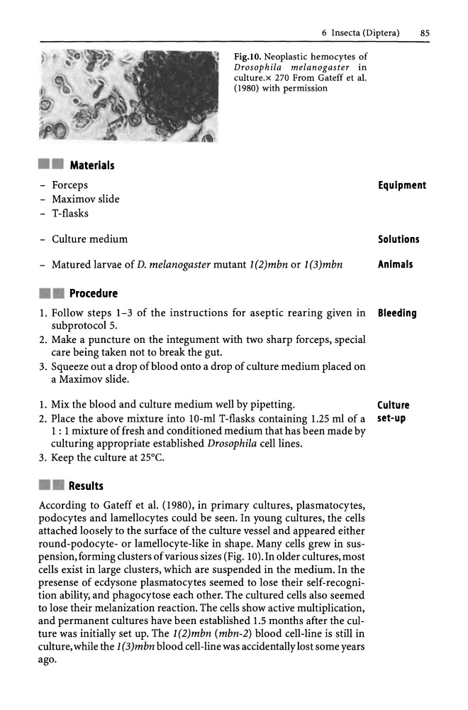
Fig.IO. Neoplastic hemocytes of
Drosophila melanogaster in
culture. x 270 From Gateff et al.
(1980) with permission
- Matured larvae of D. melanogaster mutant 1 (2)mbn or 1 (3)mbn
• • Procedure
Equipment
Solutions
Animals
1. Follow steps 1-3 of the instructions for aseptic rearing given in Bleeding
subprotocol 5.
2. Make a puncture on the integument with two sharp forceps, special
care being taken not to break the gut.
3. Squeeze out a drop of blood onto a drop of culture medium placed on
a Maximov slide.
1. Mix the blood and culture medium well by pipetting.
Culture
2. Place the above mixture into 10-ml T-flasks containing 1.25 ml of a set-up
1 : 1 mixture of fresh and conditioned medium that has been made by
culturing appropriate established Drosophila cell lines.
3. Keep the culture at 25°C.
• • Results
According to Gateff et al. (1980), in primary cultures, plasmatocytes,
podocytes and lamellocytes could be seen. In young cultures, the cells
attached loosely to the surface of the culture vessel and appeared either
round-podocyte- or lamellocyte-like in shape. Many cells grew in suspension, forming clusters of various sizes (Fig. 10). In older cultures, most
cells exist in large clusters, which are suspended in the medium. In the
presense of ecdysone plasmatocytes seemed to lose their self-recognition ability, and phagocytose each other. The cultured cells also seemed
to lose their melanization reaction. The cells show active multiplication,
and permanent cultures have been established 1.5 months after the culture was initially set up. The 1 (2)mbn (mbn-2) blood cell-line is still in
culture, while the 1 (3 )mbn blood cell-line was accidentally lost some years
ago.
85
• • Materials
- Forceps
- Maximov slide
- T-flasks
- Culture medium
Fig.IO. Neoplastic hemocytes of
Drosophila melanogaster in
culture. x 270 From Gateff et al.
(1980) with permission
- Matured larvae of D. melanogaster mutant 1 (2)mbn or 1 (3)mbn
• • Procedure
Equipment
Solutions
Animals
1. Follow steps 1-3 of the instructions for aseptic rearing given in Bleeding
subprotocol 5.
2. Make a puncture on the integument with two sharp forceps, special
care being taken not to break the gut.
3. Squeeze out a drop of blood onto a drop of culture medium placed on
a Maximov slide.
1. Mix the blood and culture medium well by pipetting.
Culture
2. Place the above mixture into 10-ml T-flasks containing 1.25 ml of a set-up
1 : 1 mixture of fresh and conditioned medium that has been made by
culturing appropriate established Drosophila cell lines.
3. Keep the culture at 25°C.
• • Results
According to Gateff et al. (1980), in primary cultures, plasmatocytes,
podocytes and lamellocytes could be seen. In young cultures, the cells
attached loosely to the surface of the culture vessel and appeared either
round-podocyte- or lamellocyte-like in shape. Many cells grew in suspension, forming clusters of various sizes (Fig. 10). In older cultures, most
cells exist in large clusters, which are suspended in the medium. In the
presense of ecdysone plasmatocytes seemed to lose their self-recognition ability, and phagocytose each other. The cultured cells also seemed
to lose their melanization reaction. The cells show active multiplication,
and permanent cultures have been established 1.5 months after the culture was initially set up. The 1 (2)mbn (mbn-2) blood cell-line is still in
culture, while the 1 (3 )mbn blood cell-line was accidentally lost some years
ago.