

6 Insecta (Diptera)
83
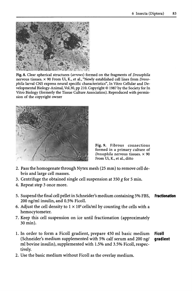
Fig. 8. Clear spherical structures (arrows) formed on the fragments of Drosophila
nervous tissues. x 90 From Ui, K., et aI., "Newly established cell lines from Drosophila larval CNS express neural specific characteristics", In Vitro Cellular and Developmental Biology-Animal, Vo1.30, pp 210. Copyright © 1987 by the Society for In
Vitro Biology (formerly the Tissue Culture Association). Reproduced with permission of the copyright owner
Fig. 9. Fibrous connections
formed in a primary culture of
Drosophila nervous tissues. x 90
From Ui, K., et aI., ditto
2. Pass the homogenate through Nytex mesh (25 mm) to remove cell debris and large cell masses.
3. Centrifuge the obtained single cell suspension at 350 g for 5 min.
4. Repeat step 3 once more.
5. Suspend the final cell pellet in Schneider's medium containing 5% FBS, Fractionation
200 ng/ml insulin, and 0.5% Ficoll.
6. Adjust the cell density to 1 x 10 6 cells/ml by counting the cells with a
hemocytometer.
7. Keep this cell suspension on ice until fractionation (approximately
30 min).
l. In order to form a Ficoll gradient, prepare 450 ml basic medium Ficoll
(Schneider's medium supplemented with 5% calf serum and 200 ng/ gradient
ml bovine insulin), supplemented with l.5% and 3.5% Ficoll, respectively.
2. Use the basic medium without Ficoll as the overlay medium.
83
Fig. 8. Clear spherical structures (arrows) formed on the fragments of Drosophila
nervous tissues. x 90 From Ui, K., et aI., "Newly established cell lines from Drosophila larval CNS express neural specific characteristics", In Vitro Cellular and Developmental Biology-Animal, Vo1.30, pp 210. Copyright © 1987 by the Society for In
Vitro Biology (formerly the Tissue Culture Association). Reproduced with permission of the copyright owner
Fig. 9. Fibrous connections
formed in a primary culture of
Drosophila nervous tissues. x 90
From Ui, K., et aI., ditto
2. Pass the homogenate through Nytex mesh (25 mm) to remove cell debris and large cell masses.
3. Centrifuge the obtained single cell suspension at 350 g for 5 min.
4. Repeat step 3 once more.
5. Suspend the final cell pellet in Schneider's medium containing 5% FBS, Fractionation
200 ng/ml insulin, and 0.5% Ficoll.
6. Adjust the cell density to 1 x 10 6 cells/ml by counting the cells with a
hemocytometer.
7. Keep this cell suspension on ice until fractionation (approximately
30 min).
l. In order to form a Ficoll gradient, prepare 450 ml basic medium Ficoll
(Schneider's medium supplemented with 5% calf serum and 200 ng/ gradient
ml bovine insulin), supplemented with l.5% and 3.5% Ficoll, respectively.
2. Use the basic medium without Ficoll as the overlay medium.