

60
Methods for Setting Up Primary Cultures Specific to Animal Groups
A ~_§~1.~
o
SUDd
,.
AdeiltioD
~
.till
W •• bllll by
of modio
Clo ••
c::::::::;::.r
liquid .boni"
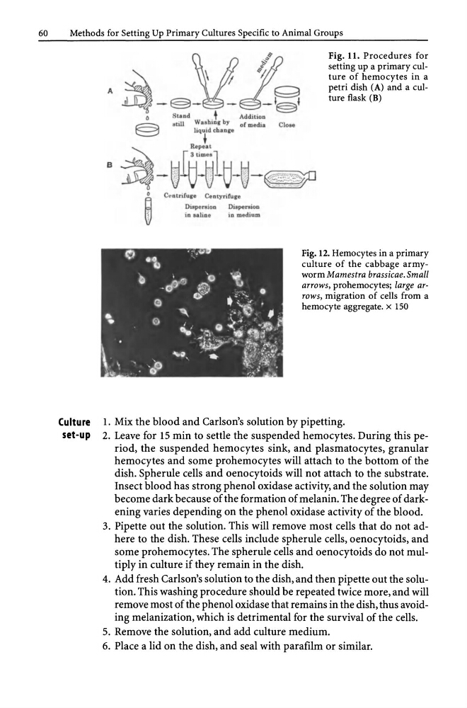
Fig. 11. Procedures for
setting up a primary culture of hemocytes in a
petri dish (A) and a culture flask (B)
S ~ 1;-:!~1
iq - ~ltj-~-~-~-~
~
0 1
CeDlnfu,o Contyrifu ••
Dj.penion
DuperaioD
in ,.liDe
in medium
Fig. 12. Hemocytes in a primary
culture of the cabbage armyworm Mamestra brassicae. Small
arrows, prohemocytes; large arrows, migration of cells from a
hemocyte aggregate. x 150
Culture 1. Mix the blood and Carlson's solution by pipetting.
set-up 2. Leave for 15 min to settle the suspended hemocytes. During this period, the suspended hemocytes sink, and plasmatocytes, granular
hemocytes and some prohemocytes will attach to the bottom of the
dish. Spherule cells and oenocytoids will not attach to the substrate.
Insect blood has strong phenol oxidase activity, and the solution may
become dark because of the formation of melanin. The degree of darkening varies depending on the phenol oxidase activity of the blood.
3. Pipette out the solution. This will remove most cells that do not adhere to the dish. These cells include spherule cells, oenocytoids, and
some prohemocytes. The spherule cells and oenocytoids do not multiply in culture if they remain in the dish.
4. Add fresh Carlson's solution to the dish, and then pipette out the solution. This washing procedure should be repeated twice more, and will
remove most of the phenol oxidase that remains in the dish, thus avoiding melanization, which is detrimental for the survival of the cells.
5. Remove the solution, and add culture medium.
6. Place a lid on the dish, and seal with parafilm or similar.
Methods for Setting Up Primary Cultures Specific to Animal Groups
A ~_§~1.~
o
SUDd
,.
AdeiltioD
~
.till
W •• bllll by
of modio
Clo ••
c::::::::;::.r
liquid .boni"
Fig. 11. Procedures for
setting up a primary culture of hemocytes in a
petri dish (A) and a culture flask (B)
S ~ 1;-:!~1
iq - ~ltj-~-~-~-~
~
0 1
CeDlnfu,o Contyrifu ••
Dj.penion
DuperaioD
in ,.liDe
in medium
Fig. 12. Hemocytes in a primary
culture of the cabbage armyworm Mamestra brassicae. Small
arrows, prohemocytes; large arrows, migration of cells from a
hemocyte aggregate. x 150
Culture 1. Mix the blood and Carlson's solution by pipetting.
set-up 2. Leave for 15 min to settle the suspended hemocytes. During this period, the suspended hemocytes sink, and plasmatocytes, granular
hemocytes and some prohemocytes will attach to the bottom of the
dish. Spherule cells and oenocytoids will not attach to the substrate.
Insect blood has strong phenol oxidase activity, and the solution may
become dark because of the formation of melanin. The degree of darkening varies depending on the phenol oxidase activity of the blood.
3. Pipette out the solution. This will remove most cells that do not adhere to the dish. These cells include spherule cells, oenocytoids, and
some prohemocytes. The spherule cells and oenocytoids do not multiply in culture if they remain in the dish.
4. Add fresh Carlson's solution to the dish, and then pipette out the solution. This washing procedure should be repeated twice more, and will
remove most of the phenol oxidase that remains in the dish, thus avoiding melanization, which is detrimental for the survival of the cells.
5. Remove the solution, and add culture medium.
6. Place a lid on the dish, and seal with parafilm or similar.