

5 Insecta (Lepidoptera)
49
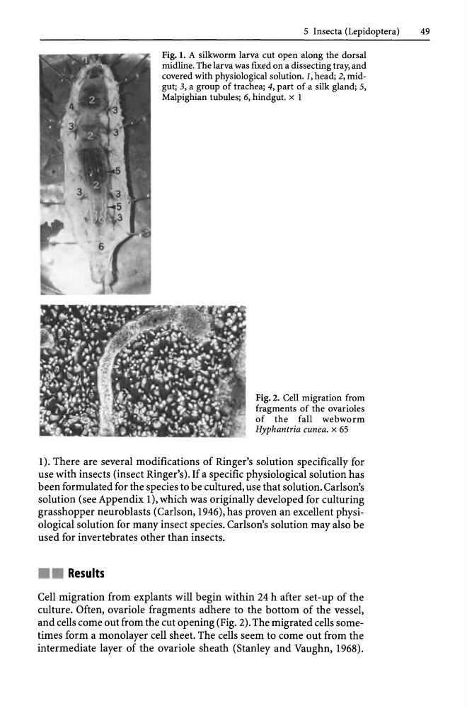
Fig. 1. A silkworm larva cut open along the dorsal
midline. The larva was fIxed on a dissecting tray, and
covered with physiological solution. 1, head; 2, midgut; 3, a group of trachea; 4, part of a silk gland; 5,
Malpighian tubules; 6, hindgut. x 1
Fig. 2. Cell migration from
fragments of the ovarioles
of the fall webworm
Hyphantria cunea. x 65
1). There are several modifications of Ringer's solution specifically for
use with insects (insect Ringer's). If a specific physiological solution has
been formulated for the species to be cultured, use that solution. Carlson's
solution (see Appendix 1), which was originally developed for culturing
grasshopper neuroblasts (Carlson, 1946), has proven an excellent physiological solution for many insect species. Carlson's solution may also be
used for invertebrates other than insects.
•
Results
Cell migration from explants will begin within 24 h after set-up of the
culture. Often, ovariole fragments adhere to the bottom of the vessel,
and cells come out from the cut opening (Fig. 2). The migrated cells sometimes form a monolayer cell sheet. The cells seem to come out from the
intermediate layer of the ovariole sheath (Stanley and Vaughn, 1968).
49
Fig. 1. A silkworm larva cut open along the dorsal
midline. The larva was fIxed on a dissecting tray, and
covered with physiological solution. 1, head; 2, midgut; 3, a group of trachea; 4, part of a silk gland; 5,
Malpighian tubules; 6, hindgut. x 1
Fig. 2. Cell migration from
fragments of the ovarioles
of the fall webworm
Hyphantria cunea. x 65
1). There are several modifications of Ringer's solution specifically for
use with insects (insect Ringer's). If a specific physiological solution has
been formulated for the species to be cultured, use that solution. Carlson's
solution (see Appendix 1), which was originally developed for culturing
grasshopper neuroblasts (Carlson, 1946), has proven an excellent physiological solution for many insect species. Carlson's solution may also be
used for invertebrates other than insects.
•
Results
Cell migration from explants will begin within 24 h after set-up of the
culture. Often, ovariole fragments adhere to the bottom of the vessel,
and cells come out from the cut opening (Fig. 2). The migrated cells sometimes form a monolayer cell sheet. The cells seem to come out from the
intermediate layer of the ovariole sheath (Stanley and Vaughn, 1968).