

308
Related Techniques
Dilution of
cell
suspension
1. Dilute the cell suspension with cloning medium, and dispense the suspension to a small petri dish (3 cm diameter) so as to make a cell
density of 1 cell/microscope field using a 10 x lO objective and ocular
lens combination.
2. Place an inverted microscope in a laminar flow cabinet after wiping
the surface of the microscope with 0.1 % benzalkonium chloride.
3. Place the petri dish on the stage of the microscope.
4. Bring the cells into the center of the field.
Capillary 1. Heat the tip of a Pasteur pipette using a small flame of a burner like a
pilot flame and draw the pipette tip out in two steps, the second step
using a still smaller flame.
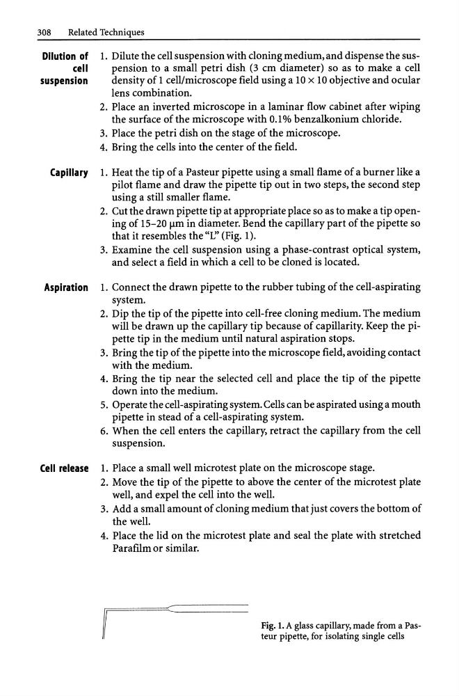
2. Cut the drawn pipette tip at appropriate place so as to make a tip opening of 15-20 !lm in diameter. Bend the capillary part of the pipette so
that it resembles the "L" (Fig. 1).
3. Examine the cell suspension using a phase-contrast optical system,
and select a field in which a cell to be cloned is located.
Aspiration 1. Connect the drawn pipette to the rubber tubing of the cell-aspirating
system.
2. Dip the tip of the pipette into cell-free cloning medium. The medium
will be drawn up the capillary tip because of capillarity. Keep the pipette tip in the medium until natural aspiration stops.
3. Bring the tip of the pipette into the microscope field, avoiding contact
with the medium.
4. Bring the tip near the selected cell and place the tip of the pipette
down into the medium.
5. Operate the cell-aspirating system. Cells can be aspirated using a mouth
pipette in stead of a cell-aspirating system.
6. When the cell enters the capillary, retract the capillary from the cell
suspension.
Cell release 1. Place a small well microtest plate on the microscope stage.
2. Move the tip of the pipette to above the center of the microtest plate
well, and expel the cell into the well.
3. Add a small amount of cloning medium that just covers the bottom of
the well.
4. Place the lid on the microtest plate and seal the plate with stretched
Parafilm or similar.
Fig. 1. A glass capillary, made from a Pasteur pipette, for isolating single cells
Related Techniques
Dilution of
cell
suspension
1. Dilute the cell suspension with cloning medium, and dispense the suspension to a small petri dish (3 cm diameter) so as to make a cell
density of 1 cell/microscope field using a 10 x lO objective and ocular
lens combination.
2. Place an inverted microscope in a laminar flow cabinet after wiping
the surface of the microscope with 0.1 % benzalkonium chloride.
3. Place the petri dish on the stage of the microscope.
4. Bring the cells into the center of the field.
Capillary 1. Heat the tip of a Pasteur pipette using a small flame of a burner like a
pilot flame and draw the pipette tip out in two steps, the second step
using a still smaller flame.
2. Cut the drawn pipette tip at appropriate place so as to make a tip opening of 15-20 !lm in diameter. Bend the capillary part of the pipette so
that it resembles the "L" (Fig. 1).
3. Examine the cell suspension using a phase-contrast optical system,
and select a field in which a cell to be cloned is located.
Aspiration 1. Connect the drawn pipette to the rubber tubing of the cell-aspirating
system.
2. Dip the tip of the pipette into cell-free cloning medium. The medium
will be drawn up the capillary tip because of capillarity. Keep the pipette tip in the medium until natural aspiration stops.
3. Bring the tip of the pipette into the microscope field, avoiding contact
with the medium.
4. Bring the tip near the selected cell and place the tip of the pipette
down into the medium.
5. Operate the cell-aspirating system. Cells can be aspirated using a mouth
pipette in stead of a cell-aspirating system.
6. When the cell enters the capillary, retract the capillary from the cell
suspension.
Cell release 1. Place a small well microtest plate on the microscope stage.
2. Move the tip of the pipette to above the center of the microtest plate
well, and expel the cell into the well.
3. Add a small amount of cloning medium that just covers the bottom of
the well.
4. Place the lid on the microtest plate and seal the plate with stretched
Parafilm or similar.
Fig. 1. A glass capillary, made from a Pasteur pipette, for isolating single cells