

294
Organ Culture
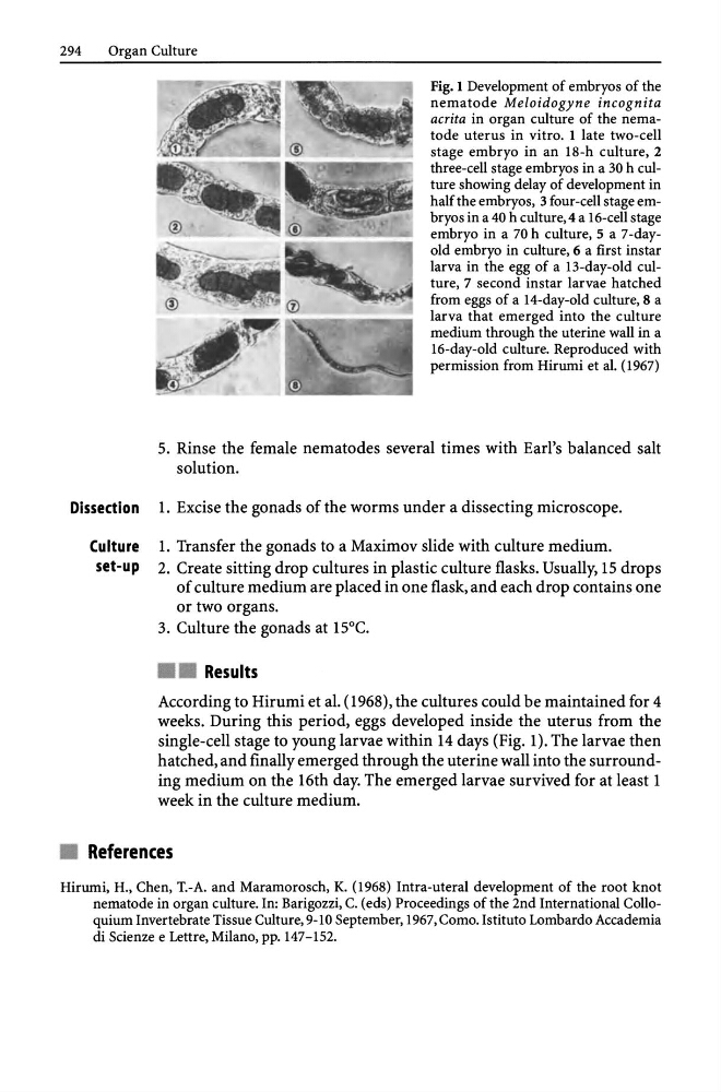
Fig. 1 Development of embryos of the
nematode Meloidogyne incognita
acrita in organ culture of the nematode uterus in vitro. 1 late two-cell
stage embryo in an 18-h culture, 2
three-cell stage embryos in a 30 h culture showing delay of development in
half the embryos, 3 four-cell stage embryos in a 40 h culture, 4 a 16-cell stage
embryo in a 70 h culture,S a 7-dayold embryo in culture, 6 a first ins tar
larva in the egg of a 13-day-old culture, 7 second ins tar larvae hatched
from eggs of a 14-day-old culture, 8 a
larva that emerged into the culture
medium through the uterine wall in a
16-day-old culture. Reproduced with
permission from Hirumi et al. (1967)
5. Rinse the female nematodes several times with Earl's balanced salt
solution.
Dissection 1. Excise the gonads of the worms under a dissecting microscope.
Culture 1. Transfer the gonads to a Maximov slide with culture medium.
set-up 2. Create sitting drop cultures in plastic culture flasks. Usually, 15 drops
of culture medium are placed in one flask, and each drop contains one
or two organs.
3. Culture the gonads at 15°C.
• • Results
According to Hirumi et al. (l968), the cultures could be maintained for 4
weeks. During this period, eggs developed inside the uterus from the
single-cell stage to young larvae within 14 days (Fig. O. The larvae then
hatched, and finally emerged through the uterine wall into the surrounding medium on the 16th day. The emerged larvae survived for at least 1
week in the culture medium.
• References
Hirumi, H., Chen, T.-A. and Maramorosch, K. (1968) Intra-uteral development of the root knot
nematode in organ culture. In: Barigozzi, C. (eds) Proceedings of the 2nd International Colloquium Invertebrate Tissue Culture, 9-10 September, 1967, Como. Istituto Lombardo Accademia
di Scienze e Lettre, Milano, pp. 147-152.
Organ Culture
Fig. 1 Development of embryos of the
nematode Meloidogyne incognita
acrita in organ culture of the nematode uterus in vitro. 1 late two-cell
stage embryo in an 18-h culture, 2
three-cell stage embryos in a 30 h culture showing delay of development in
half the embryos, 3 four-cell stage embryos in a 40 h culture, 4 a 16-cell stage
embryo in a 70 h culture,S a 7-dayold embryo in culture, 6 a first ins tar
larva in the egg of a 13-day-old culture, 7 second ins tar larvae hatched
from eggs of a 14-day-old culture, 8 a
larva that emerged into the culture
medium through the uterine wall in a
16-day-old culture. Reproduced with
permission from Hirumi et al. (1967)
5. Rinse the female nematodes several times with Earl's balanced salt
solution.
Dissection 1. Excise the gonads of the worms under a dissecting microscope.
Culture 1. Transfer the gonads to a Maximov slide with culture medium.
set-up 2. Create sitting drop cultures in plastic culture flasks. Usually, 15 drops
of culture medium are placed in one flask, and each drop contains one
or two organs.
3. Culture the gonads at 15°C.
• • Results
According to Hirumi et al. (l968), the cultures could be maintained for 4
weeks. During this period, eggs developed inside the uterus from the
single-cell stage to young larvae within 14 days (Fig. O. The larvae then
hatched, and finally emerged through the uterine wall into the surrounding medium on the 16th day. The emerged larvae survived for at least 1
week in the culture medium.
• References
Hirumi, H., Chen, T.-A. and Maramorosch, K. (1968) Intra-uteral development of the root knot
nematode in organ culture. In: Barigozzi, C. (eds) Proceedings of the 2nd International Colloquium Invertebrate Tissue Culture, 9-10 September, 1967, Como. Istituto Lombardo Accademia
di Scienze e Lettre, Milano, pp. 147-152.