

214
Methods for Setting Up Primary Cultures Specific to Animal Groups
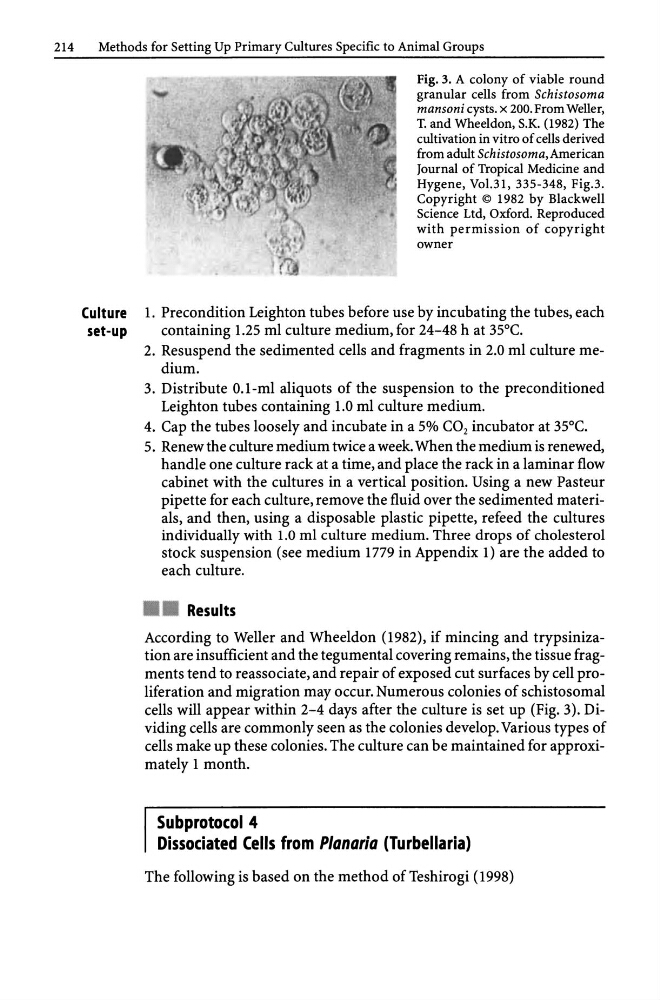
Fig. 3. A colony of viable round
granular cells from Schistosoma
mansoni cysts. x 200. From Weller,
T. and Wheeldon, S.K. (1982) The
cultivation in vitro of cells derived
from adult Schistosoma,American
Journal of Tropical Medicine and
Hygene, Vol.31, 335-348, Fig.3.
Copyright © 1982 by Blackwell
Science Ltd, Oxford. Reproduced
with permission of copyright
owner
Culture 1. Precondition Leighton tubes before use by incubating the tubes, each
set-up
containing 1.25 ml culture medium, for 24-48 h at 35°C.
2. Resuspend the sedimented cells and fragments in 2.0 ml culture medium.
3. Distribute O.I-ml aliquots of the suspension to the preconditioned
Leighton tubes containing 1.0 ml culture medium.
4. Cap the tubes loosely and incubate in a 5% CO 2 incubator at 35°C.
5. Renew the culture medium twice a week. When the medium is renewed,
handle one culture rack at a time, and place the rack in a laminar flow
cabinet with the cultures in a vertical position. Using a new Pasteur
pipette for each culture, remove the fluid over the sedimented materials, and then, using a disposable plastic pipette, refeed the cultures
individually with 1.0 ml culture medium. Three drops of cholesterol
stock suspension (see medium 1779 in Appendix 1) are the added to
each culture .
• • Results
According to Weller and Wheeldon (1982), if mincing and trypsinization are insufficient and the tegumental covering remains, the tissue fragments tend to reassociate, and repair of exposed cut surfaces by cell proliferation and migration may occur. Numerous colonies of schistosomal
cells will appear within 2-4 days after the culture is set up (Fig. 3). Dividing cells are commonly seen as the colonies develop. Various types of
cells make up these colonies. The culture can be maintained for approximately 1 month.
Subprotocol 4
Dissociated Cells from P/anaria (Turbellaria)
The following is based on the method of Teshirogi (1998)
Methods for Setting Up Primary Cultures Specific to Animal Groups
Fig. 3. A colony of viable round
granular cells from Schistosoma
mansoni cysts. x 200. From Weller,
T. and Wheeldon, S.K. (1982) The
cultivation in vitro of cells derived
from adult Schistosoma,American
Journal of Tropical Medicine and
Hygene, Vol.31, 335-348, Fig.3.
Copyright © 1982 by Blackwell
Science Ltd, Oxford. Reproduced
with permission of copyright
owner
Culture 1. Precondition Leighton tubes before use by incubating the tubes, each
set-up
containing 1.25 ml culture medium, for 24-48 h at 35°C.
2. Resuspend the sedimented cells and fragments in 2.0 ml culture medium.
3. Distribute O.I-ml aliquots of the suspension to the preconditioned
Leighton tubes containing 1.0 ml culture medium.
4. Cap the tubes loosely and incubate in a 5% CO 2 incubator at 35°C.
5. Renew the culture medium twice a week. When the medium is renewed,
handle one culture rack at a time, and place the rack in a laminar flow
cabinet with the cultures in a vertical position. Using a new Pasteur
pipette for each culture, remove the fluid over the sedimented materials, and then, using a disposable plastic pipette, refeed the cultures
individually with 1.0 ml culture medium. Three drops of cholesterol
stock suspension (see medium 1779 in Appendix 1) are the added to
each culture .
• • Results
According to Weller and Wheeldon (1982), if mincing and trypsinization are insufficient and the tegumental covering remains, the tissue fragments tend to reassociate, and repair of exposed cut surfaces by cell proliferation and migration may occur. Numerous colonies of schistosomal
cells will appear within 2-4 days after the culture is set up (Fig. 3). Dividing cells are commonly seen as the colonies develop. Various types of
cells make up these colonies. The culture can be maintained for approximately 1 month.
Subprotocol 4
Dissociated Cells from P/anaria (Turbellaria)
The following is based on the method of Teshirogi (1998)