

200
Methods for Setting Up Primary Cultures Specific to Animal Groups
sup. 9
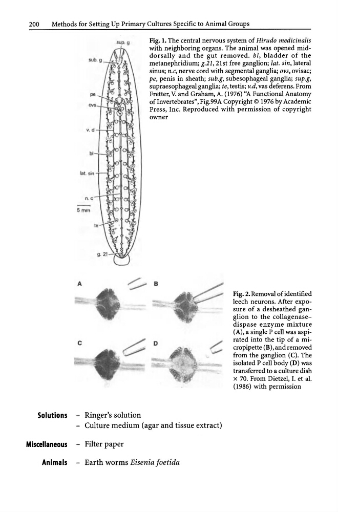
Fig. 1. The central nervous system of Hirudo medicinalis
with neighboring organs. The animal was opened middorsally and the gut removed. bl, bladder of the
metanephridium; g.21, 21st free ganglion; lat. sin, lateral
sinus; n.c, nerve cord with segmental ganglia; ovs, ovisac;
pe, penis in sheath; sub.g, subesophageal ganglia; sup.g,
supraesophageal ganglia; te, testis; v.d, vas deferens. From
Fretter, V. and Graham, A. (1976) "A Functional Anatomy
of Invertebrates", Fig.99A Copyright © 1976 by Academic
Press, Inc. Reproduced with permission of copyright
owner
A
~ B
D
Solutions - Ringer's solution
- Culture medium (agar and tissue extract)
Miscellaneous - Filter paper
Animals - Earth worms Eisenia foetida
Fig. 2. Removal of identified
leech neurons. After exposure of a desheathed ganglion to the collagenasedispase enzyme mixture
(A), a single P cell was aspirated into the tip of a micropipette (B),and removed
from the ganglion (C). The
isolated P cell body (D) was
transferred to a culture dish
x 70. From Dietzel, I. et al.
(1986) with permission
Methods for Setting Up Primary Cultures Specific to Animal Groups
sup. 9
Fig. 1. The central nervous system of Hirudo medicinalis
with neighboring organs. The animal was opened middorsally and the gut removed. bl, bladder of the
metanephridium; g.21, 21st free ganglion; lat. sin, lateral
sinus; n.c, nerve cord with segmental ganglia; ovs, ovisac;
pe, penis in sheath; sub.g, subesophageal ganglia; sup.g,
supraesophageal ganglia; te, testis; v.d, vas deferens. From
Fretter, V. and Graham, A. (1976) "A Functional Anatomy
of Invertebrates", Fig.99A Copyright © 1976 by Academic
Press, Inc. Reproduced with permission of copyright
owner
A
~ B
D
Solutions - Ringer's solution
- Culture medium (agar and tissue extract)
Miscellaneous - Filter paper
Animals - Earth worms Eisenia foetida
Fig. 2. Removal of identified
leech neurons. After exposure of a desheathed ganglion to the collagenasedispase enzyme mixture
(A), a single P cell was aspirated into the tip of a micropipette (B),and removed
from the ganglion (C). The
isolated P cell body (D) was
transferred to a culture dish
x 70. From Dietzel, I. et al.
(1986) with permission