

15 Mollusca
189
17
16
14
,,'...;::~t----'<-- 5
7
11
Fig. 21. Inner structure of a nudibranch
(Doris) . 1, Central ganglia; 2, lymph gland; 3,
osmeterium; 4, gonad; 5, liver; 6, ventricle; 7,
rectum; 8, auricle; 9, nephrostome; 10, excretory pore; 11, anus; 12, periproct branchia; 13,
branchial vein; 14, mantle vein; 15, liver; 16,
intestine; 17, esophagus; 18, stomach; 19, salivary gland. From Pelseneer, P., 1906
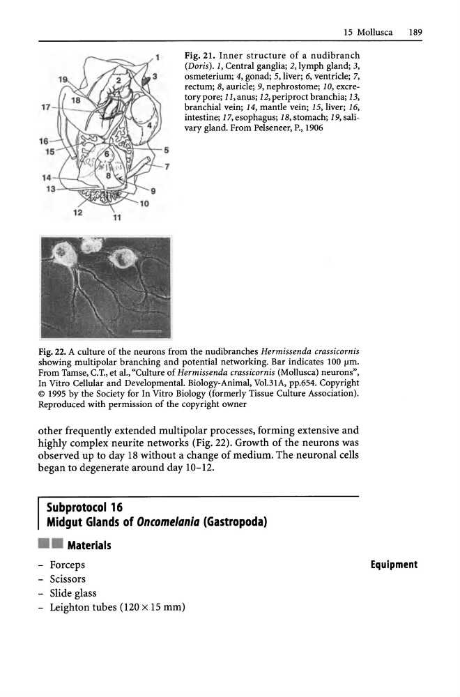
Fig. 22. A culture of the neurons from the nudibranches Hermissenda crassicornis
showing multipolar branching and potential networking. Bar indicates 100 flm.
From Tamse, C.T., et al., "Culture of Hermissenda crassicornis (Mollusca) neurons",
In Vitro Cellular and Developmental. Biology-Animal, Vo1.31A, pp.654. Copyright
© 1995 by the Society for In Vitro Biology (formerly Tissue Culture Association).
Reproduced with permission of the copyright owner
other frequently extended multipolar processes, forming extensive and
highly complex neurite networks (Fig. 22). Growth of the neurons was
observed up to day 18 without a change of medium. The neuronal cells
began to degenerate around day 1O-l2.
Subprotocol 16
Midgut Glands of Oncomelania (Gastropoda)
• • Materials
- Forceps
- Scissors
- Slide glass
- Leighton tubes (120 x 15 mm)
Equipment
189
17
16
14
,,'...;::~t----'<-- 5
7
11
Fig. 21. Inner structure of a nudibranch
(Doris) . 1, Central ganglia; 2, lymph gland; 3,
osmeterium; 4, gonad; 5, liver; 6, ventricle; 7,
rectum; 8, auricle; 9, nephrostome; 10, excretory pore; 11, anus; 12, periproct branchia; 13,
branchial vein; 14, mantle vein; 15, liver; 16,
intestine; 17, esophagus; 18, stomach; 19, salivary gland. From Pelseneer, P., 1906
Fig. 22. A culture of the neurons from the nudibranches Hermissenda crassicornis
showing multipolar branching and potential networking. Bar indicates 100 flm.
From Tamse, C.T., et al., "Culture of Hermissenda crassicornis (Mollusca) neurons",
In Vitro Cellular and Developmental. Biology-Animal, Vo1.31A, pp.654. Copyright
© 1995 by the Society for In Vitro Biology (formerly Tissue Culture Association).
Reproduced with permission of the copyright owner
other frequently extended multipolar processes, forming extensive and
highly complex neurite networks (Fig. 22). Growth of the neurons was
observed up to day 18 without a change of medium. The neuronal cells
began to degenerate around day 1O-l2.
Subprotocol 16
Midgut Glands of Oncomelania (Gastropoda)
• • Materials
- Forceps
- Scissors
- Slide glass
- Leighton tubes (120 x 15 mm)
Equipment