

15 Mollusca
187
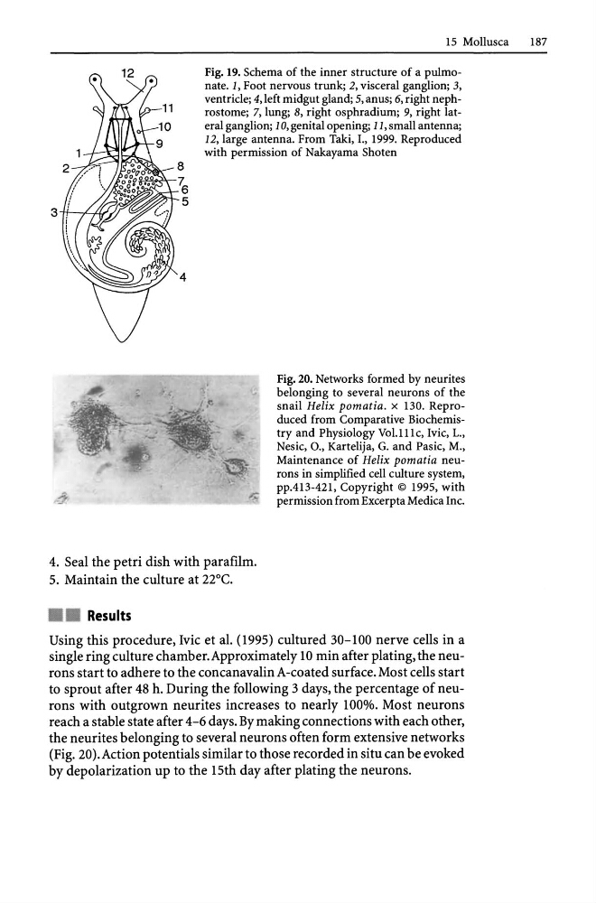
Fig. 19. Schema of the inner structure of a pulmonate. 1, Foot nervous trunk; 2, visceral ganglion; 3,
ventricle; 4, left midgut gland; 5, anus; 6, right nephrostome; 7, lung; 8, right osphradium; 9, right lateral ganglion; 10, genital opening; 11, small antenna;
12, large antenna. From Taki, I., 1999. Reproduced
with permission of Nakayama Shoten
Fig. 20. Networks formed by neurites
belonging to several neurons of the
snail Helix pomatia. x 130. Reproduced from Comparative Biochemistry and Physiology Vol.lll c, Ivic, 1.,
Nesic, 0., Kartelija, G. and Pasic, M.,
Maintenance of Helix pomatia neurons in simplified cell culture system,
ppA13-421, Copyright © 1995, with
permission from Excerpta Medica Inc.
4. Seal the petri dish with parafilm.
5. Maintain the culture at 22°C.
• • Results
Using this procedure, Ivic et al. (1995) cultured 30-100 nerve cells in a
single ring culture chamber. Approximately 10 min after plating, the neurons start to adhere to the concanavalin A-coated surface. Most cells start
to sprout after 48 h. During the following 3 days, the percentage of neurons with outgrown neurites increases to nearly 100%. Most neurons
reach a stable state after 4-6 days. By making connections with each other,
the neurites belonging to several neurons often form extensive networks
(Fig. 20).Action potentials similar to those recorded in situ can be evoked
by depolarization up to the 15th day after plating the neurons.
187
Fig. 19. Schema of the inner structure of a pulmonate. 1, Foot nervous trunk; 2, visceral ganglion; 3,
ventricle; 4, left midgut gland; 5, anus; 6, right nephrostome; 7, lung; 8, right osphradium; 9, right lateral ganglion; 10, genital opening; 11, small antenna;
12, large antenna. From Taki, I., 1999. Reproduced
with permission of Nakayama Shoten
Fig. 20. Networks formed by neurites
belonging to several neurons of the
snail Helix pomatia. x 130. Reproduced from Comparative Biochemistry and Physiology Vol.lll c, Ivic, 1.,
Nesic, 0., Kartelija, G. and Pasic, M.,
Maintenance of Helix pomatia neurons in simplified cell culture system,
ppA13-421, Copyright © 1995, with
permission from Excerpta Medica Inc.
4. Seal the petri dish with parafilm.
5. Maintain the culture at 22°C.
• • Results
Using this procedure, Ivic et al. (1995) cultured 30-100 nerve cells in a
single ring culture chamber. Approximately 10 min after plating, the neurons start to adhere to the concanavalin A-coated surface. Most cells start
to sprout after 48 h. During the following 3 days, the percentage of neurons with outgrown neurites increases to nearly 100%. Most neurons
reach a stable state after 4-6 days. By making connections with each other,
the neurites belonging to several neurons often form extensive networks
(Fig. 20).Action potentials similar to those recorded in situ can be evoked
by depolarization up to the 15th day after plating the neurons.