

15 Mollusca
185
2. Transfer the tissue to 0.025% potassium permanganate.
3. Sterilize the tissue in 1 % (v/v) Clorox for 1 min.
4. Rinse the tissues in double-strength L-15 medium four times.
1. Chop the tissues into I-mm 3 pieces using scissors.
Dissociation
2. Digest the tissue fragments with a 0.25% (v/v) solution of trypsinEDTA in 2.6% (w/v) NaCI solution for 12 h.
3. Dissociate the cells by vigorous pipetting.
1. Inoculate approximately 2 x 10 7 dissociated cells into a 25-cm 2 plastic Culture
flask containing double-strength L-15 medium fortified with 20% (vi set-up
v) hard clam hemolymph, 5% (v/v) FBS, 100 IV/ml penicillin, and
100 Jlg/ml streptomycin.
2. Culture at 24°C by renewing the culture medium every week.
• • Comments
To obtain clam hemolymph, insert a needle through a notch cut on the
bivalve and suck the hemolymph out.
• • Results
Cell migration occurs from the explanted gill fragments. The migrated
cells multiply by mitosis. These cells do not adhere to the substrate, and
multiply in suspension. In cell cultures of R. decussatus, cells migrate
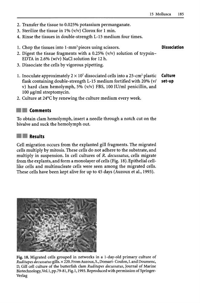
from the explants, and form a monolayer of cells (Fig. 18). Epithelial celllike cells and multinucleate cells were seen among the migrated cells.
These cells have been kept alive for up to 45 days (AuZQux et aI., 1993).
Fig. 18. Migrated cells grouped in networks in a I-day-old primary culture of
Ruditapes decussatus gills. x 220. From Auzoux, S., Domart - Coulon, I. and Doumenc,
D, Gill cell culture of the butterfish clam Ruditapes decussatus, Journal of Marine
Biotechnology, VoU, pp.79-81, Fig.I, 1993. Reproduced with permission of SpringerVerlag
185
2. Transfer the tissue to 0.025% potassium permanganate.
3. Sterilize the tissue in 1 % (v/v) Clorox for 1 min.
4. Rinse the tissues in double-strength L-15 medium four times.
1. Chop the tissues into I-mm 3 pieces using scissors.
Dissociation
2. Digest the tissue fragments with a 0.25% (v/v) solution of trypsinEDTA in 2.6% (w/v) NaCI solution for 12 h.
3. Dissociate the cells by vigorous pipetting.
1. Inoculate approximately 2 x 10 7 dissociated cells into a 25-cm 2 plastic Culture
flask containing double-strength L-15 medium fortified with 20% (vi set-up
v) hard clam hemolymph, 5% (v/v) FBS, 100 IV/ml penicillin, and
100 Jlg/ml streptomycin.
2. Culture at 24°C by renewing the culture medium every week.
• • Comments
To obtain clam hemolymph, insert a needle through a notch cut on the
bivalve and suck the hemolymph out.
• • Results
Cell migration occurs from the explanted gill fragments. The migrated
cells multiply by mitosis. These cells do not adhere to the substrate, and
multiply in suspension. In cell cultures of R. decussatus, cells migrate
from the explants, and form a monolayer of cells (Fig. 18). Epithelial celllike cells and multinucleate cells were seen among the migrated cells.
These cells have been kept alive for up to 45 days (AuZQux et aI., 1993).
Fig. 18. Migrated cells grouped in networks in a I-day-old primary culture of
Ruditapes decussatus gills. x 220. From Auzoux, S., Domart - Coulon, I. and Doumenc,
D, Gill cell culture of the butterfish clam Ruditapes decussatus, Journal of Marine
Biotechnology, VoU, pp.79-81, Fig.I, 1993. Reproduced with permission of SpringerVerlag