

182
Methods for Setting Up Primary Cultures Specific to Animal Groups
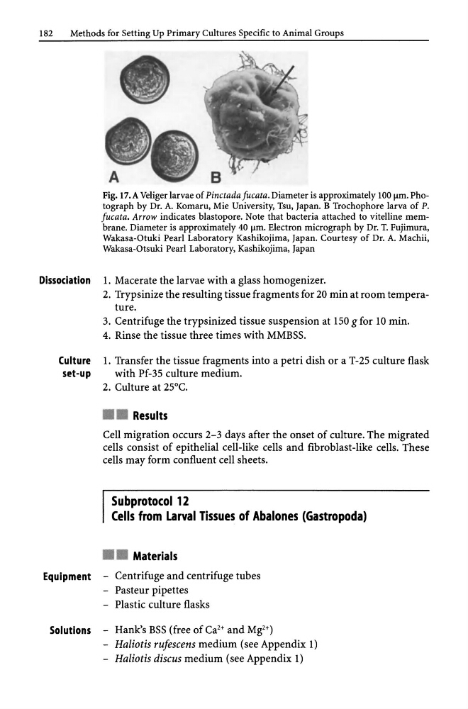
Fig. 17.A Veliger larvae of Pinctada Jucata. Diameter is approximately 100 flm. Photograph by Dr. A. Komaru, Mie University, Tsu, Japan. B Trochophore larva of P.
Jucata. Arrow indicates blastopore. Note that bacteria attached to vitelline membrane. Diameter is approximately 40 flm. Electron micrograph by Dr. T. Fujimura,
Wakasa-Otuki Pearl Laboratory Kashikojima, Japan. Courtesy of Dr. A. Machii,
Wakasa-Otsuki Pearl Laboratory, Kashikojima, Japan
Dissociation 1. Macerate the larvae with a glass homogenizer.
2. Trypsinize the resulting tissue fragments for 20 min at room temperature.
3. Centrifuge the trypsinized tissue suspension at 150 g for 10 min.
4. Rinse the tissue three times with MMBSS.
Culture 1. Transfer the tissue fragments into a petri dish or a T-25 culture flask
set-up
with Pf-35 culture medium.
2. Culture at 25°C .
• • Results
Cell migration occurs 2-3 days after the onset of culture. The migrated
cells consist of epithelial cell-like cells and fibroblast-like cells. These
cells may form confluent cell sheets.
Subprotocol 12
Cells from Larval Tissues of Abalones (Gastropoda)
• • Materials
Equipment - Centrifuge and centrifuge tubes
- Pasteur pipettes
- Plastic culture flasks
Solutions - Hank's BSS (free of Ca 2 + and Mg2+)
- Haliotis rufescens medium (see Appendix 1)
- Haliotis discus medium (see Appendix 1)
Methods for Setting Up Primary Cultures Specific to Animal Groups
Fig. 17.A Veliger larvae of Pinctada Jucata. Diameter is approximately 100 flm. Photograph by Dr. A. Komaru, Mie University, Tsu, Japan. B Trochophore larva of P.
Jucata. Arrow indicates blastopore. Note that bacteria attached to vitelline membrane. Diameter is approximately 40 flm. Electron micrograph by Dr. T. Fujimura,
Wakasa-Otuki Pearl Laboratory Kashikojima, Japan. Courtesy of Dr. A. Machii,
Wakasa-Otsuki Pearl Laboratory, Kashikojima, Japan
Dissociation 1. Macerate the larvae with a glass homogenizer.
2. Trypsinize the resulting tissue fragments for 20 min at room temperature.
3. Centrifuge the trypsinized tissue suspension at 150 g for 10 min.
4. Rinse the tissue three times with MMBSS.
Culture 1. Transfer the tissue fragments into a petri dish or a T-25 culture flask
set-up
with Pf-35 culture medium.
2. Culture at 25°C .
• • Results
Cell migration occurs 2-3 days after the onset of culture. The migrated
cells consist of epithelial cell-like cells and fibroblast-like cells. These
cells may form confluent cell sheets.
Subprotocol 12
Cells from Larval Tissues of Abalones (Gastropoda)
• • Materials
Equipment - Centrifuge and centrifuge tubes
- Pasteur pipettes
- Plastic culture flasks
Solutions - Hank's BSS (free of Ca 2 + and Mg2+)
- Haliotis rufescens medium (see Appendix 1)
- Haliotis discus medium (see Appendix 1)