

178
Methods for Setting Up Primary Cultures Specific to Animal Groups
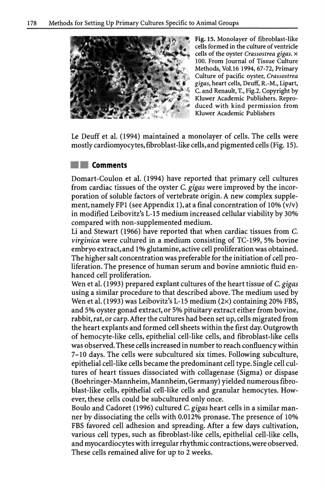
Fig. 15. Monolayer of fibroblast-like
cells formed in the culture of ventricle
cells of the oyster Crassostrea gigas. x
100. From Journal of Tissue Culture
Methods, Vo1.l6 1994,67-72, Primary
Culture of pacific oyster, Crassostrea
gigas, heart cells, Deuff, R.-M., Lipart,
C. and Renault, T., Fig.2. Copyright by
Kluwer Academic Publishers. Reproduced with kind permission from
Kluwer Academic Publishers
Le Deuff et al. (1994) maintained a monolayer of cells. The cells were
mostly cardiomyocytes,fibroblast-like cells, and pigmented cells (Fig. 15).
Comments
Domart-Coulon et al. (1994) have reported that primary cell cultures
from cardiac tissues of the oyster C. gigas were improved by the incorporation of soluble factors of vertebrate origin. A new complex supplement,namely FPl (see Appendix I), at a final concentration of 10% (v/v)
in modified Leibovitz's L-15 medium increased cellular viability by 30%
compared with non-supplemented medium.
Li and Stewart (1966) have reported that when cardiac tissues from C.
virginica were cultured in a medium consisting of TC-199, 5% bovine
embryo extract, and 1 % glutamine, active cell proliferation was obtained.
The higher salt concentration was preferable for the initiation of cell proliferation. The presence of human serum and bovine amniotic fluid enhanced cell proliferation.
Wen et al. (1993) prepared explant cultures of the heart tissue of C. gigas
using a similar procedure to that described above. The medium used by
Wen et al. (1993) was Leibovitz's L-15 medium (2x) containing 20% FBS,
and 5% oyster gonad extract, or 5% pituitary extract either from bovine,
rabbit, rat, or carp. After the cultures had been set up, cells migrated from
the heart explants and formed cell sheets within the first day. Outgrowth
of hemocyte-like cells, epithelial cell-like cells, and fibroblast-like cells
was observed. These cells increased in number to reach confluency within
7 -10 days. The cells were subcultured six times. Following subculture,
epithelial cell-like cells became the predominant cell type. Single cell cultures of heart tissues dissociated with collagenase (Sigma) or dispase
(Boehringer-Mannheim, Mannheim, Germany) yielded numerous fibroblast-like cells, epithelial cell-like cells and granular hemocytes. However, these cells could be subcultured only once.
Boulo and Cadoret (1996) cultured C. gigas heart cells in a similar manner by dissociating the cells with 0.012% pronase. The presence of 10%
FBS favored cell adhesion and spreading. After a few days cultivation,
various cell types, such as fibroblast-like cells, epithelial cell-like cells,
and myocardiocytes with irregular rhythmic contractions, were observed.
These cells remained alive for up to 2 weeks.
Methods for Setting Up Primary Cultures Specific to Animal Groups
Fig. 15. Monolayer of fibroblast-like
cells formed in the culture of ventricle
cells of the oyster Crassostrea gigas. x
100. From Journal of Tissue Culture
Methods, Vo1.l6 1994,67-72, Primary
Culture of pacific oyster, Crassostrea
gigas, heart cells, Deuff, R.-M., Lipart,
C. and Renault, T., Fig.2. Copyright by
Kluwer Academic Publishers. Reproduced with kind permission from
Kluwer Academic Publishers
Le Deuff et al. (1994) maintained a monolayer of cells. The cells were
mostly cardiomyocytes,fibroblast-like cells, and pigmented cells (Fig. 15).
Comments
Domart-Coulon et al. (1994) have reported that primary cell cultures
from cardiac tissues of the oyster C. gigas were improved by the incorporation of soluble factors of vertebrate origin. A new complex supplement,namely FPl (see Appendix I), at a final concentration of 10% (v/v)
in modified Leibovitz's L-15 medium increased cellular viability by 30%
compared with non-supplemented medium.
Li and Stewart (1966) have reported that when cardiac tissues from C.
virginica were cultured in a medium consisting of TC-199, 5% bovine
embryo extract, and 1 % glutamine, active cell proliferation was obtained.
The higher salt concentration was preferable for the initiation of cell proliferation. The presence of human serum and bovine amniotic fluid enhanced cell proliferation.
Wen et al. (1993) prepared explant cultures of the heart tissue of C. gigas
using a similar procedure to that described above. The medium used by
Wen et al. (1993) was Leibovitz's L-15 medium (2x) containing 20% FBS,
and 5% oyster gonad extract, or 5% pituitary extract either from bovine,
rabbit, rat, or carp. After the cultures had been set up, cells migrated from
the heart explants and formed cell sheets within the first day. Outgrowth
of hemocyte-like cells, epithelial cell-like cells, and fibroblast-like cells
was observed. These cells increased in number to reach confluency within
7 -10 days. The cells were subcultured six times. Following subculture,
epithelial cell-like cells became the predominant cell type. Single cell cultures of heart tissues dissociated with collagenase (Sigma) or dispase
(Boehringer-Mannheim, Mannheim, Germany) yielded numerous fibroblast-like cells, epithelial cell-like cells and granular hemocytes. However, these cells could be subcultured only once.
Boulo and Cadoret (1996) cultured C. gigas heart cells in a similar manner by dissociating the cells with 0.012% pronase. The presence of 10%
FBS favored cell adhesion and spreading. After a few days cultivation,
various cell types, such as fibroblast-like cells, epithelial cell-like cells,
and myocardiocytes with irregular rhythmic contractions, were observed.
These cells remained alive for up to 2 weeks.