

15 Mollusca
175
radular
.heath
gm
""':S;:~_ operculum
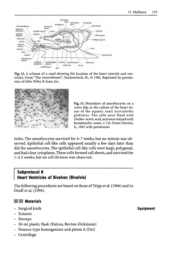
Fig. 12. A schema of a snail showing the location of the heart (auricle and ventricle). From "The Invertebrates", Stachowitsch, M., © 1992. Reprinted by permission oOohn Wiley & Sons, Inc.
Fig. 13. Monolayer of amoebocytes on a
cover slip, in the culture of the heart tissue of the aquatic snail Australorbis
glabratus. The cells were fixed with
Zenker-acetic acid, and were stained with
hematoxylin-eosin. x 125. From Chernin,
E., 1963 with permission
tides. The amoebocytes survived for 4-7 weeks, but no mitosis was observed. Epithelial cell-like cells appeared usually a few days later than
did the amoebocytes. The epithelial cell-like cells were large, polygonal,
and had dear cytoplasm. These cells formed cell sheets, and survived for
1-2.5 weeks, but no cell division was observed.
Subprotocol 8
Heart Ventricles of Bivalves (Bivalvia)
The following procedures are based on those of Tripp et al. (1966) and Le
Deuff et al. (1994) .
• • Materials
- Surgical knife
- Scissors
- Forceps
- 30-ml plastic flask (Falcon, Becton-Dickinson)
- Dounce-type homogenizer and piston A (Osi)
- Centrifuge
Equipment
175
radular
.heath
gm
""':S;:~_ operculum
Fig. 12. A schema of a snail showing the location of the heart (auricle and ventricle). From "The Invertebrates", Stachowitsch, M., © 1992. Reprinted by permission oOohn Wiley & Sons, Inc.
Fig. 13. Monolayer of amoebocytes on a
cover slip, in the culture of the heart tissue of the aquatic snail Australorbis
glabratus. The cells were fixed with
Zenker-acetic acid, and were stained with
hematoxylin-eosin. x 125. From Chernin,
E., 1963 with permission
tides. The amoebocytes survived for 4-7 weeks, but no mitosis was observed. Epithelial cell-like cells appeared usually a few days later than
did the amoebocytes. The epithelial cell-like cells were large, polygonal,
and had dear cytoplasm. These cells formed cell sheets, and survived for
1-2.5 weeks, but no cell division was observed.
Subprotocol 8
Heart Ventricles of Bivalves (Bivalvia)
The following procedures are based on those of Tripp et al. (1966) and Le
Deuff et al. (1994) .
• • Materials
- Surgical knife
- Scissors
- Forceps
- 30-ml plastic flask (Falcon, Becton-Dickinson)
- Dounce-type homogenizer and piston A (Osi)
- Centrifuge
Equipment