

15 Mollusca
173
15
1 -::==+:::.:::1::2 -=4=1;--4~
3
-
4 - - .
5
6 - "U"loo~"'"
7
11
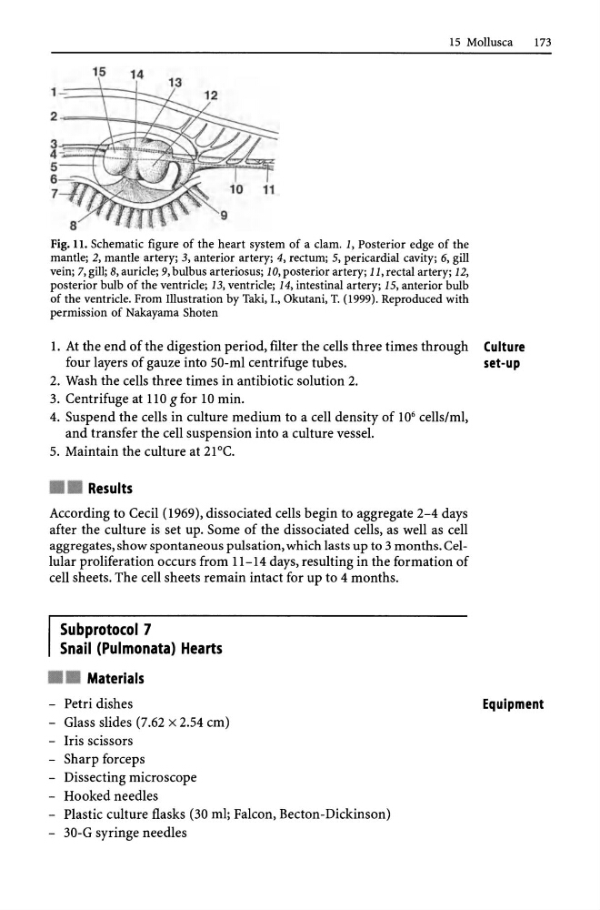
Fig. 11. Schematic figure of the heart system of a clam. 1, Posterior edge of the
mantle; 2, mantle artery; 3, anterior artery; 4, rectum; 5, pericardial cavity; 6, gill
vein; 7, gill; 8, auricle; 9, bulbus arteriosus; 10, posterior artery; 11, rectal artery; 12,
posterior bulb of the ventricle; 13, ventricle; 14, intestinal artery; 15, anterior bulb
of the ventricle. From Illustration by rald, I., Okutani, r. (1999). Reproduced with
permission of Nakayama Shoten
1. At the end of the digestion period, filter the cells three times through Culture
four layers of gauze into 50-ml centrifuge tubes.
set-up
2. Wash the cells three times in antibiotic solution 2.
3. Centrifuge at 110 gfor 10 min.
4. Suspend the cells in culture medium to a cell density of 10 6 cells/ml,
and transfer the cell suspension into a culture vessel.
5. Maintain the culture at 21°C.
• • Results
According to Cecil (1969), dissociated cells begin to aggregate 2-4 days
after the culture is set up. Some of the dissociated cells, as well as cell
aggregates, show spontaneous pulsation, which lasts up to 3 months. Cellular proliferation occurs from 11-14 days, resulting in the formation of
cell sheets. The cell sheets remain intact for up to 4 months.
Subprotocol 7
Snail (Pulmonata) Hearts
• • Materials
- Petri dishes
- Glass slides (7.62 x 2.54 em)
- Iris scissors
- Sharp forceps
- Dissecting microscope
- Hooked needles
- Plastic culture flasks (30 ml; Falcon, Becton-Dickinson)
- 30-G syringe needles
Equipment
173
15
1 -::==+:::.:::1::2 -=4=1;--4~
3
-
4 - - .
5
6 - "U"loo~"'"
7
11
Fig. 11. Schematic figure of the heart system of a clam. 1, Posterior edge of the
mantle; 2, mantle artery; 3, anterior artery; 4, rectum; 5, pericardial cavity; 6, gill
vein; 7, gill; 8, auricle; 9, bulbus arteriosus; 10, posterior artery; 11, rectal artery; 12,
posterior bulb of the ventricle; 13, ventricle; 14, intestinal artery; 15, anterior bulb
of the ventricle. From Illustration by rald, I., Okutani, r. (1999). Reproduced with
permission of Nakayama Shoten
1. At the end of the digestion period, filter the cells three times through Culture
four layers of gauze into 50-ml centrifuge tubes.
set-up
2. Wash the cells three times in antibiotic solution 2.
3. Centrifuge at 110 gfor 10 min.
4. Suspend the cells in culture medium to a cell density of 10 6 cells/ml,
and transfer the cell suspension into a culture vessel.
5. Maintain the culture at 21°C.
• • Results
According to Cecil (1969), dissociated cells begin to aggregate 2-4 days
after the culture is set up. Some of the dissociated cells, as well as cell
aggregates, show spontaneous pulsation, which lasts up to 3 months. Cellular proliferation occurs from 11-14 days, resulting in the formation of
cell sheets. The cell sheets remain intact for up to 4 months.
Subprotocol 7
Snail (Pulmonata) Hearts
• • Materials
- Petri dishes
- Glass slides (7.62 x 2.54 em)
- Iris scissors
- Sharp forceps
- Dissecting microscope
- Hooked needles
- Plastic culture flasks (30 ml; Falcon, Becton-Dickinson)
- 30-G syringe needles
Equipment